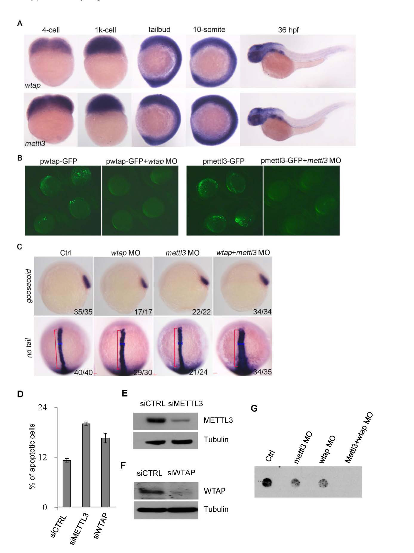
Fig. S6
Expression pattern and functional assay of WTAP and METTL3 in zebrafish embryos (A) Whole-mount in situ hybridization (WISH) shows ubiquitous expression of WTAP and METTL3 during embryogenesis, respectively. (B) To test the efficiency of WTAP- and METTL3-MOs one-cell embryos were injected with 25 pg of pWTAP-GFP plasmid DNA alone or in combination with WTAP MO (left panel) or METTL3 MO (right panel). (C) To test the expression of the neuro-ectoderm marker “goosecoid” and mesoderm marker “no tail” in embryos injected with individual or a combination of WTAP and METTL3 MOs. Red bracket indicates the length and blue bracket the width of “no tail” expression in the notochord. (D) HeLa cells were treated with siControl, siMETTL3 or siWTAP RNAi for 48h, then stained with annexin V-FITC and PI and subjected to apoptotic analyses by flow cytometry. (E-F) Western blots showing knock down efficiency of siMETTL3 (E) and siWTAP (F) in cells used in the apoptotic assay shown in (D). (G) Zebrafish embryos were treated with control, WTAP- and METTL3-MOs as indicated. mRNA was extracted and 50 ng of mRNA was spotted onto a nylon membrane and m6A levels were detected with anti-m6A antibody.

