
Fig. 3
- ID
- ZDB-IMAGE-140404-3
- Publication
- Gray et al., 2014 - Loss of col8a1a function during zebrafish embryogenesis results in congenital vertebral malformations
- All Figures
- Figures for Gray et al., 2014
|
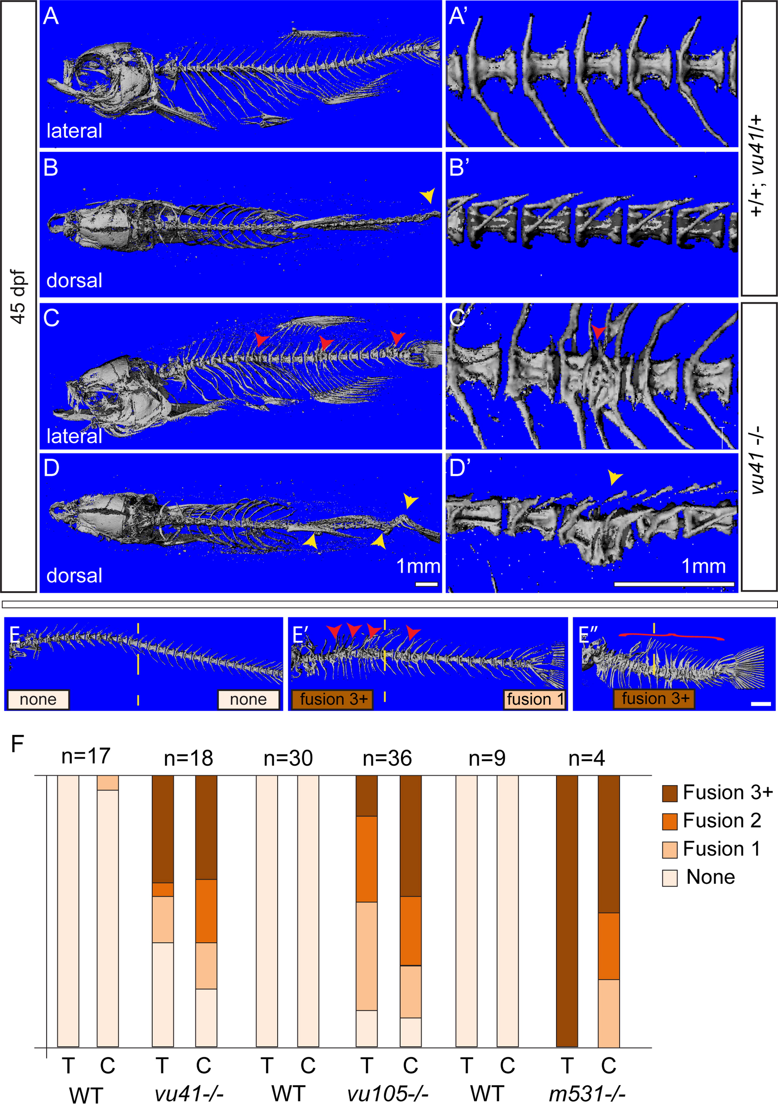
Fig. 3 Homozyogus col8a1a mutant adults display vertebral fusions and scoliosis. MicroCT imaging of WT (A–B′) and col8a1a mutant zebrafish (C–D′) adults (45dpf) (A–E′′) in both lateral views (A, C, E–E′′) with insets (A′ and C′) and dorsal views (B and D) with insets (B′, D′) (A, A′, and B′) WT vertebrae with one neural and hemal arches per vertebral body. (B) Representative scoliosis in the tail of a WT adult (yellow arrowhead). (C and C′) Representative levvu41-/- zebrafish with VM (red arrowheads). (D and D′) Representative levvu41-/- multiple scoliosis along the axis (yellow arrowheads) and (E–E′′) Representative individuals scored for the presence of individual VM (red arrowheads) or complete fusion of vertebral column (red bracket) in the thoracic or caudal regions (as number of fused regions). (F) Graphed categorical data of VM from all lev alleles and unaffected WT siblings in thoracic (T) and caudal (C) regions. All scale bars (A–D), (A′–D′), and (E–E′′)=1 mM.
Reprinted from Developmental Biology, 386(1), Gray, R.S., Wilm, T.P., Smith, J., Bagnat, M., Dale, R.M., Topczewski, J., Johnson, S.L., and Solnica-Krezel, L., Loss of col8a1a function during zebrafish embryogenesis results in congenital vertebral malformations, 72-85, Copyright (2014) with permission from Elsevier. Full text @ Dev. Biol.