
Fig. S7
- ID
- ZDB-IMAGE-140325-23
- Publication
- Wang et al., 2014 - Notch3 establishes brain vascular integrity by regulating pericyte number
- All Figures
- Figures for Wang et al., 2014
|
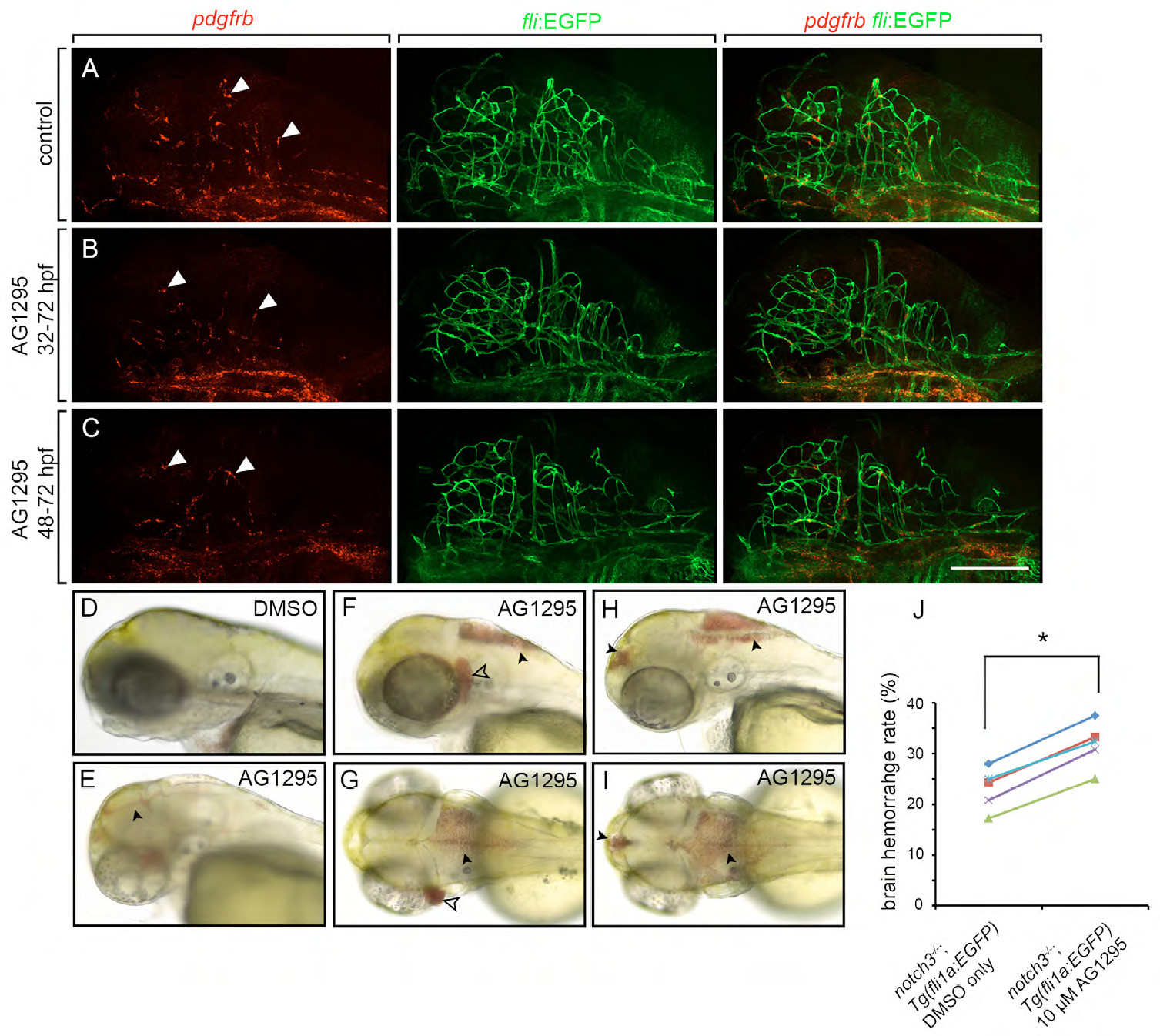
Fig. S7
Inhibition of Pdgfr-β activity produces a pericyte deficit and brain hemorrahge. (A) pdgfrb+ brain pericytes in DMSO treated control larvae. (B,C) Larvae treated with 25 μM AG1295 from 32-72 hpf, and from 48-72 hpf, respectively, showed a reduction in pdgfrb+ brain pericyte number. Arrowheads, pdgfrb+ brain pericytes. Scale bar, 200 μm. (A-C) All images show confocal projections of whole brains, viewed from lateral. (D-I) Brain hemorrhage in AG1295 treated zebrafish larvae. (D) DMSO treated control at 3 dpf. (F-I) 25μm AG1295 treated larva at 3 dpf. (E,F,H) Lateral views. (G,I) Dorsal views of (F,H), respectively. (J) notch3fh332 homozygous mutants showed a more penetrant hemorrhage phenotype when treated with 10 μM AG1295 (n=5, *P<0.05, by paired t-test). Arrowheads, blood pooling at the brain ventricle; open arrowhead, blood pooling behind the eye.