
Fig. 6
- ID
- ZDB-IMAGE-140320-2
- Genes
- Publication
- Schmid et al., 2000 - Equivalent genetic roles for bmp7/snailhouse and bmp2b/swirl in dorsoventral pattern formation
- All Figures
- Figures for Schmid et al., 2000
|
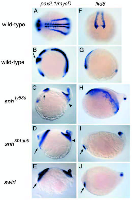
Fig. 6 In situ hybridizations with pax2.1 and myoD (A-E) and fkd6 (F-J) in wild-type, snh/bmp7 and swrta72/bmp2b mutant embryos at the 5-somite stage. Lateral views, anterior to the left, except (A,F). (A) The somitic expression of myoD and the pronephric expression of pax2.1 in a dorsal-posterior view of a wild-type embryo. (B) The midbrain/hindbrain boundary (MHB) expression of pax2.1 (arrow) in a wild-type embryo. (C) snhty68a displays a slight lateral expansion of the MHB (arrow), not visible in this lateral view. The somites posterior to somite 2 encircle the embryo (arrowhead). (D) In snhsb1aub mutant embryos the MHB (arrow) and all somites (arrowhead) encircle the embryo. (E) Homozygous swrta72 mutants are indistinguishable from snhsb1aub mutants. fkd6 expression in the cranial neural crest progenitors in wild-type in an anterior (F) (dorsal to the top) and lateral (G) view. fkd6 expression is also observed in the tail bud (G). (H) Expanded cranial neural crest expression in snhty68a. In snhsb1aub (I) and swr/bmp2b (J) mutants, the number of cranial neural crest progenitors is severely reduced (arrow).