
Fig. 5
- ID
- ZDB-IMAGE-140313-7
- Publication
- Renucci et al., 1996 - An activated form of type I serine/threonine kinase receptor TARAM-A reveals a specific signalling pathway involved in fish head organiser formation
- All Figures
- Figures for Renucci et al., 1996
|
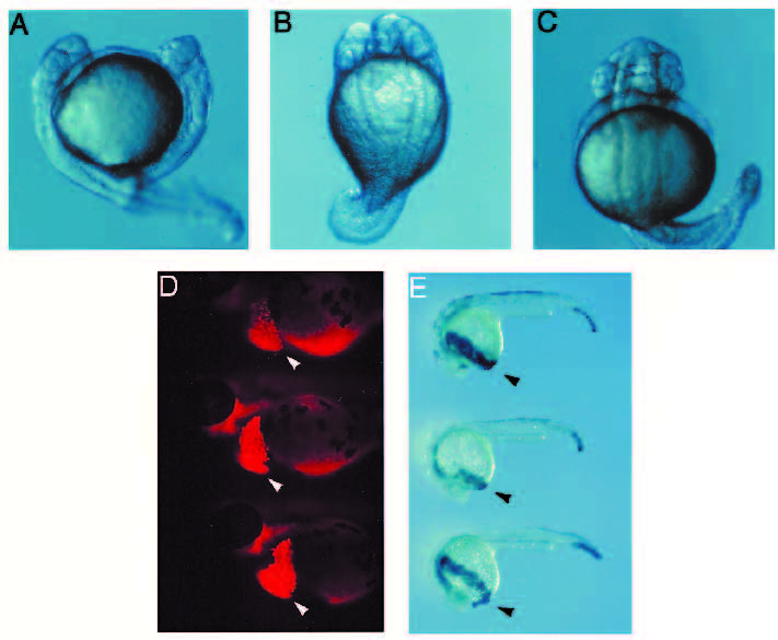
Fig. 5 Duplication of anterior structures in TARAM-A-D-injected embryos. (A-C) TARAM-A-D RNA was injected at 0.1 mg/ml into a marginal cell from 16-cellstage embryos. Embryos are observed after 24 hours, three specimens with duplication of anterior structures are shown. (A) A complete duplication of the head is observed, the two heads are facing each other (side view). (B) The two heads are contiguous (dorsal view). (C) One additional eye in a frontal position is observed. (D,E) Fate of TARAMA- D-expressing cells using coinjection of the rhodamine dextran lineage tracer (D), or the b-gal RNA tracer (40 μg/ml) (E), into one marginal cell of a 16-cell-stage embryo. (D) Left side view from the head and trunk of three 48-hour embryos (anterior to the left). The hatching gland (arrowheads) and head mesendoderm are strongly fluorescent. The apparent labelling of the yolk ball is due to light refraction. (E) Left side view from three 24-hour embryos stained for β-gal activity. Hatching glands (arrowheads) and head mesendoderm display strong β-gal activity. Some scattered periderm cells as well as undifferentiated tailbud cells are also labelled.