
|
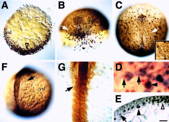
Fig. 7 Fkd2 protein is an early marker of endodermal precursors. (A) Dome stage, animal pole view; presumptive dorsal side (open arrowhead). (B) 60% epiboly, dorsal oblique view; presumptive endoderm precursor cells in focus on the left side (arrow) and larger, more irregular-shaped yolk syncytial nuclei in focus on the right side. (C) 90% epiboly, dorsal view; the lateral wing of endoderm precursor cells (arrow). Inset is a closeup of this location showing smaller endoderm nuclei, and larger yolk cell nuclei. (D) 90% epiboly, higher magnification showing characteristic endoderm precursor cells (arrows) labeled in vivo with fixable lineage tracer and then fixed and stained for the tracer (blue) and Fkd2 immunoreactivity (brown); 23 cells were analyzed in this manner. Arrowhead indicates a Fkd2-positive yolk cell nucleus. (E) 90% epiboly, transverse section (10 μm) through the midline (open arrowhead), showing one of several cell nuclei in the endodermal layer (arrow) and an underlying yolk cell nucleus (arrowhead). (F) 10-somite stage, dorsal oblique view; endodermal cells in focus on the right side (arrow). (G) 18-somite stage, dorsal view; endodermal cell sheet (arrow) in midline. Scale bar: 100 μm (A-C, F), 50 μm (G, and inset C), 20 μm (E), 10 μm (D); abbreviations: nc, notochord; pcp, prechordal plate; sh, shield; ysl, yolk syncytial nuclei.