
Fig. 1
- ID
- ZDB-IMAGE-140313-33
- Genes
- Antibodies
- Publication
- Chandrasekhar et al., 1999 - The zebrafish detour gene is essential for cranial but not spinal motor neuron induction
- All Figures
- Figures for Chandrasekhar et al., 1999
|
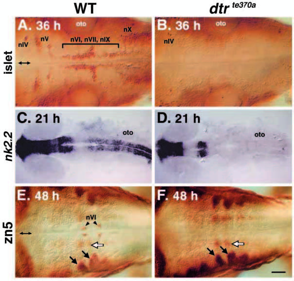
Fig. 1 Cranial motor neurons are missing in dtrte370a embryos. All panels depict dorsal views, with rostral to the left, of the hindbrain of whole-mounted embryos analyzed either by islet (A,B) or zn5 (E,F) immunohistochemistry, or by nk2.2 in situ hybridization (C,D). Double arrows (A,E) mark the midline. (A) In a 36 HPF wild-type sibling, the islet antibody labels the trigeminal motor (nV) neurons in r2 and r3, the abducens (nVI), the facial motor (nVII) and the glossopharyngeal motor (nIX) neurons in r4, r5, r6 and r7, and the vagal motor (nX) neurons in the caudal hindbrain. The antibody also labels the presumptive trochlear (nIV) neurons in r1. (B) In a dtrte370a homozygote, all cranial motor neurons, except the putative nIV neurons, are missing. (C) In a 21 HPF wild-type sibling, nk2.2 is expressed in the ventral CNS throughout the forebrain, the rostral midbrain and the hindbrain. (D) In a dtrte370a homozygote, nk2.2 expression is missing throughout the hindbrain and in the rostralmost midbrain. (E) In a 48 HPF wild-type sibling, the zn5 antibody labels the abducens neurons (nVI) in r5 and r6, and some unidentified cells just laterally (white arrow). The labeled cells located most laterally (black arrows) are the hindbrain commissural neurons. (F) In a dtrte370a homozygote, the nVI neurons are missing. However, the hindbrain commissural neurons (black arrows) and the unidentified zn5-labeled cells (white arrow) are unaffected. oto, otocyst. Scale bar, 40 μm (A,B,E,F), 100 μm (C,D).