
Image
|
Figure Caption
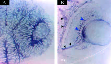
Fig. 5 Sections of in situ hybridizations using the Nrf probe in 24 hour (A) and 48 hour (B) retinae. Note that at 24 hpf, expression of nrf is detected in every cell, whereas at 48 hpf, expression is highest in the ganglion cell layer (large blue arrowheads), optic nerve and optic tracts (small black arrowheads), while very low in the PR layer.
Figure Data
Acknowledgments
This image is the copyrighted work of the attributed author or publisher, and
ZFIN has permission only to display this image to its users.
Additional permissions should be obtained from the applicable author or publisher of the image.
Full text @ Development