
|
Fig. 6
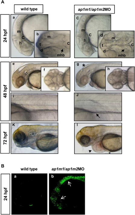
Morphological defects and apoptosis in ap1m1/ap1m2MO double morphants. A: Two-cell stage embryos were injected with 0,6 pmol/embryo of ap1m1MO plus 0,4 pmol/embryo of ap1m2MO. At 24 hpf, double morphants (c; control a) showed brain malformations with a disorganization of the midbrain–hindbrain boundary. At 48 hpf, the ventricular cavity is reduced (g,h; forebrain ventricle: black asterisk). The proximal tubule appeared irregular and not clearly defined than in wild-type embryos (i, j arrow). At 72 hpf, in double morphants the edema (arrowhead) is more pronounced (controls k,l). a, c, e, g, i, j, k, l: lateral view, anterior to the left; b,d,f,h: dorsal view. m, midbrain.; t, telencephalon; d, diencephalons; mhb, midbrain–hindbrain boundary; c, cerebellum. B: Wild-type (a) and ap1m1/ap1m2MO (b) were incubated at 24 hpf with acridine orange (AO) and then analyzed by laser confocal microscopy. Apoptotic cells are detected in the CNS of double morphants.