
Fig. 2
- ID
- ZDB-IMAGE-140303-24
- Genes
- Publication
- Moens et al., 1996 - valentino, a zebrafish gene required for normal hindbrain segmentation
- All Figures
- Figures for Moens et al., 1996
|
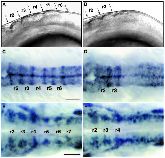
Fig. 2 val- embryos lack segment boundaries and segmental patterns of neuronal differentiation posterior to rhombomere 4. (A,B) Lateral view of live 18 h wild-type (A) and val- (B) embryos. Anterior is to the left. In val- embryos, there are no visible rhombomere boundaries posterior to the r3-r4 boundary. (C,D) Dorsal views of RNA in situ hybridizations of wild-type (C) and val- (D) embryos at 18 h showing expression of mariposa in the rhombomere boundaries. No expression is observed posterior to the r3-r4 boundary in val- embryos. (E,F) Dorsal views of RNA in situ hybridizations of wildtype (E) and val- (F) embryos at 24 h showing expression of gap43 in clusters of early differentiating neurons laterally in each rhombomere. In val- embryos, this segmental pattern of gap43 staining is lost posterior to r4. This disrupted pattern of neuronal differentiation is also observed in val- embryos stained with the HNK-1 antibody (data not shown; Metcalfe et al., 1990; Trevarrow et al., 1990). Scale bars, 50 μm.