
|
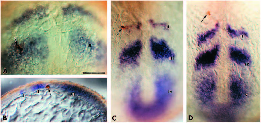
Fig. 4 Expression domains correspond to somitic primordia. (A) An embryo injected (three brown cells at the arrow) in expression domain II at 100% epiboly. This embryo is one of the control cases and was processed for in situ hybridization and anti-fluorescein antibody staining immediately after injection. (B) A parasagittal section (~100 μm) of a 3- somite stage embryo that had been injected in domain II at 100% epiboly. The arrow points to a labelled cell caudally in domain II. (C) A double-stained 6-somite embryo (whole mount) following an injection in domain II at 100% epiboly. Labelled cells are located within domain II, immediately posterior to the 6th somite (no clearly visible). (D) A double-stained 7-somite stage (whole mount) following an injection in domain II at 100% epiboly. Labelled cells are located within the 7th somite. I-V, expression domains. Scale bar, 100 μm.