
Fig. 5
- ID
- ZDB-IMAGE-140228-36
- Publication
- Stainier et al., 1995 - cloche, an early acting zebrafish gene, is required by both the endothelial and hematopoietic lineages
- All Figures
- Figures for Stainier et al., 1995
|
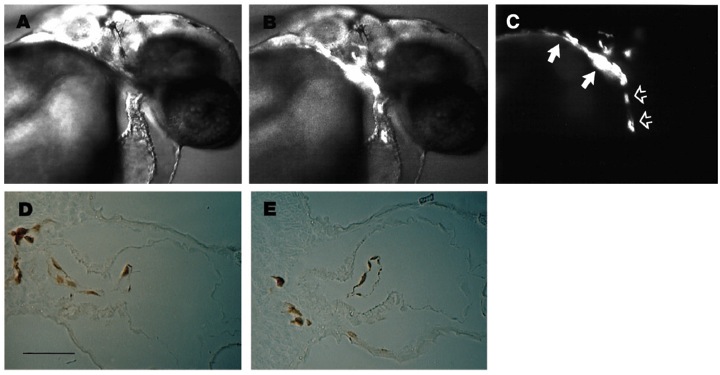
Fig. 5 Wild-type cells can form endocardium in clo mutant embryos. Nomarski (A), Nomarski + fluorescence (B), and fluorescence (C) micrographs of the same living 36 hour mutant host embryo. Labeled cells, derived from a wild-type donor embryo, populate the outflow tract (filled arrows) and form an elongated mass of tissue in the ventricular chamber of the myocardial tube (open arrows). Transverse sections through the heart of another mutant host after horseradish peroxidase staining of the biotin dextran labeled wild-type cells (D,E) reveal that transplanted wild-type cells form a distinctly elongated cell layer, the endocardial layer, inside the heart of the host embryo. When endocardium was observed in wild-type (WT) to mutant (M) transplants, all the endocardial cells were derived from WT as assessed by immunoreactivity to biotin (n=10). Scale bar 50 μm (D,E).