
Fig. 3
- ID
- ZDB-IMAGE-140221-5
- Genes
- Publication
- Drummond et al., 1998 - Early development of the zebrafish pronephros and analysis of mutations affecting pronephric function
- All Figures
- Figures for Drummond et al., 1998
|
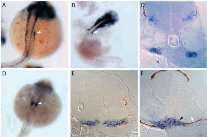
Fig. 3 Expression of pax2.1 and wt1 in the forming pronephros. (A) Whole-mount in situ hybridization shows pax2.1 is expressed in the anterior pronephric ducts (arrow) at 24 h as well as in the spinal cord. (B) At 30-32 h, a new domain of pax2.1 expression is observed anterior and medial to the pronephric ducts (arrow), in the position of the future pronephric tubules. (C) Histological sections of pax2.1 expression in the presumptive tubule primordium cells show that these cells constitute the lateral half of the nephron primordium and that the medial halves of the nephron primordium are negative. (D) Whole-mount in situ hybridization shows wt1 expression in the paired nephron primordia at 24h (arrow). (E) At 30-32 h, wt1 is uniformly expressed throughout the nephron primordia. (F) By 36 h, wt1 expression becomes restricted to a mass of cells at the midline in the position of the future glomerulus (black arrow). Lateral cells within the forming nephron in the position of the future tubule do not express wt1 (white arrow).