
Fig. 7
- ID
- ZDB-IMAGE-140117-32
- Publication
- Bitomsky et al., 2013 - Autophosphorylation and Pin1 binding coordinate DNA damage-induced HIPK2 activation and cell death
- All Figures
- Figures for Bitomsky et al., 2013
|
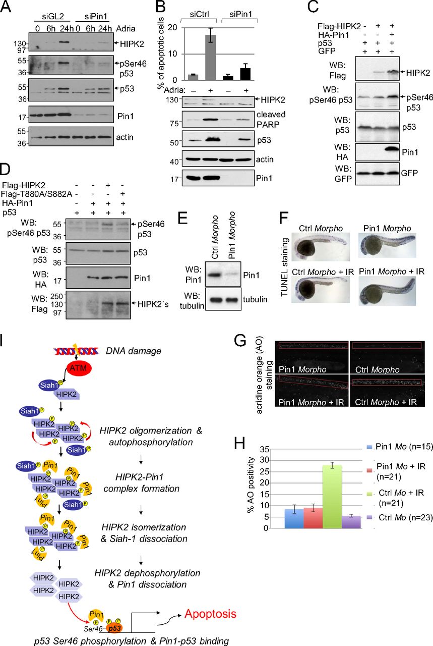
Fig. 7
Pin1 regulates HIPK2-mediated p53 Ser46 phosphorylation in human cells and is required for DNA damage-induced apoptosis in zebrafish. (A) Pin1 knockdown inhibits Adriamycin-induced HIPK2 stabilization and p53 Ser46 phosphorylation. HCT116 cells were transfected with control (GL2, luciferase) or Pin1-specific siRNA as indicated and treated with 1 μg/mL Adriamycin. Cells were lysed at the timepoints indicated and analyzed by immunoblotting. (B) Pin1 depletion impairs apoptosis induction. HCT116 cells were transfected with Pin1-specific or control siRNAs. Cells were treated 24 h with 1 μg/mL Adriamycin. Sub-G1 DNA content analysis was performed by propidium-iodide staining and FACS analysis. The graph shows means and SD from three independent experiments. Knockdown efficiency and apoptosis induction was controlled by immunoblotting. (C) Pin1 potentiates HIPK2-mediated p53 Ser46 phosphorylation. H1299 cells were transfected as indicated. Cell lysates were analyzed by immunoblotting. (D) Pin1–HIPK2 interaction seems to be important for induction of p53 Ser46 phosphorylation by Pin1. H1299 cells were transfected with the expression vectors indicated. Amounts of transfected HIPK2 expression vectors were adjusted to result in a comparable HIPK2 expression. Cell lysates were analyzed by immunoblotting. (E–H) Pin1 depletion in zebrafish embryos results in decreased apoptosis induction upon IR in vivo. (E) Zebrafish eggs were injected with Pin1-specific or control morpholinos. Knockdown of endogenous Pin1 was controlled by immunoblotting of whole-body embryo lysates. (F and G) At 24 h after fertilization (hpf) embryos were exposed to 12.5 Gy IR and analyzed 30 hpf for apoptosis induction by using either TUNEL (F) or acridine orange (AO) (G) staining. Representative images are shown. (H) Quantification of the AO staining in the zebrafish embryos. (A–G) Representative experiments are shown. (I) Proposed mechanism of HIPK2 activation upon DNA damage. For details, see Discussion.