
Fig. 10
- ID
- ZDB-IMAGE-140113-13
- Genes
- Publication
- de Oliveira-Carlos et al., 2013 - Notch receptor expression in neurogenic regions of the adult zebrafish brain
- All Figures
- Figures for de Oliveira-Carlos et al., 2013
|
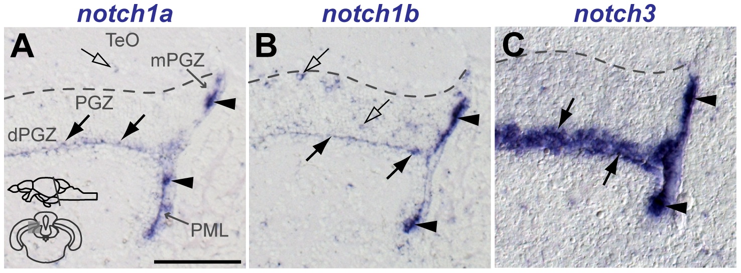
Fig. 10
Notch receptor expression in the adult zebrafish optic tectum.
Cross-sections at the indicated level through the mesencephalon; optic tectum area shown in the micrographs is indicated in the cross section schematic in A. Brighfield images show the expression (by ISH) of A, notch1a, B, notch1b and C, notch3 along the PML and mPGZ (filled arrowheads) and in the dPGZ (filled arrows). A few notch1a and notch1b cells are also found in more superficial layers of the PGZ and TeO (unfilled arrows). Abbreviations: PGZ, periventricular gray zone of the optic tectum; dPGZ, deep layer of the PGZ; mPGZ, mitotic region of the PGZ; PML, posterior mesencephalic lamina; TeO, optic tectum. Scale bars = 100 μm in A (applies to all).