
Fig. 6
- ID
- ZDB-IMAGE-131213-64
- Genes
- Publication
- Graham et al., 2013 - Epidermal keratinocyte polarity and motility require Ca2+ influx through TRPV1
- All Figures
- Figures for Graham et al., 2013
|
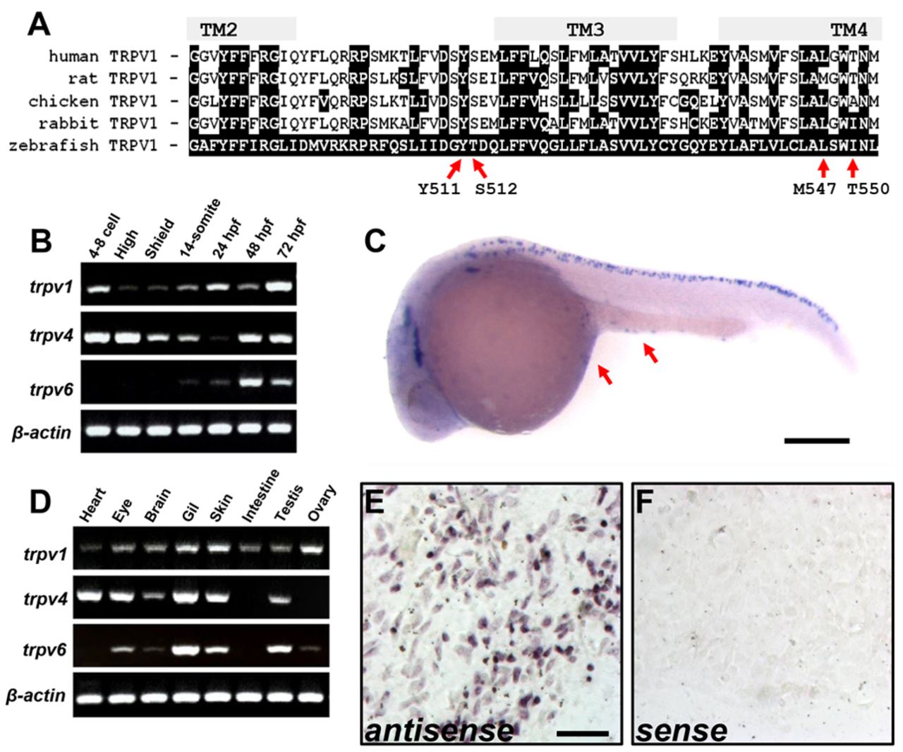
Fig. 6 Zebrafish TRPV1 is expressed in the epidermal epithelium. (A) Sequence alignment of TRPV1 proteins from different species comparing the critical residues for vanilloid activation. Defined transmembrane regions (TM) are based on the predicted membrane topology of zebrafish TRPV1. Red arrows indicate residues important for capsaicin sensitivity and are listed according to the residue positions of rat TRPV1. (B) Gene expression of TRPV members during different stages of development. (C) In situ hybridization for trpv1 in an embryo 24hours post fertilization (hpf). Arrows indicate staining in the epidermal epithelium. Scale bar: 250μm. (D) Tissue-specific gene expression of TRPV members. (E,F) In situ hybridization of the motile fraction of an explant of adult epidermal epithelia for trpv1 using antisense- and sense RNA probes. Scale bar: 50 μm.