
Fig. 4
- ID
- ZDB-IMAGE-131211-38
- Publication
- Phng et al., 2013 - Filopodia are dispensable for endothelial tip cell guidance
- All Figures
- Figures for Phng et al., 2013
|
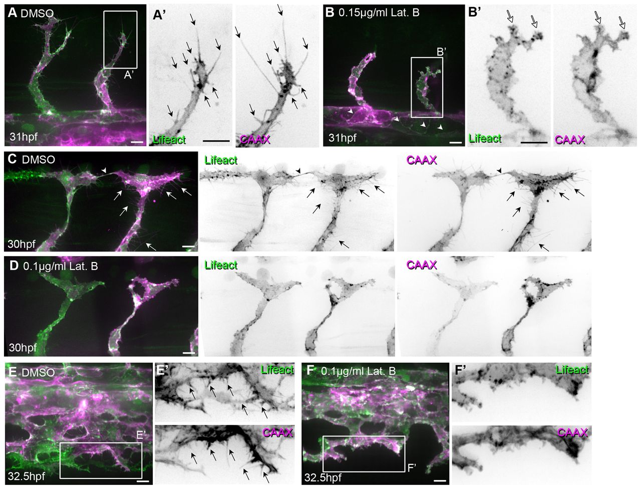
Fig. 4 Low concentrations of Latrunculin B inhibit filopodia formation. Tg(Fli1ep:Lifeact-EGFP); Tg(Kdr-l:ras-Cherry)s916 zebrafish embryos were treated with 0.4% DMSO (control) or low concentrations of Latrunculin B (Lat. B) from (A-B′) 24 to 29 hpf, (C-D′) 26 to 29 hpf or (E-F′) 26.5 to 32 hpf. (A-B′) Arrows highlight filopodia on sprouting ISVs. White arrows indicate lamellipodia-like protrusions. Arrowheads point to junctional F-actin at dorsal aorta. (C-D′) Arrows indicate filopodia. Arrowhead highlights filopodia-based contacts between two tip cells that form the DLAV. (E-F′) Arrows indicate filopodia at vein plexus. Scale bars: 10 μm.