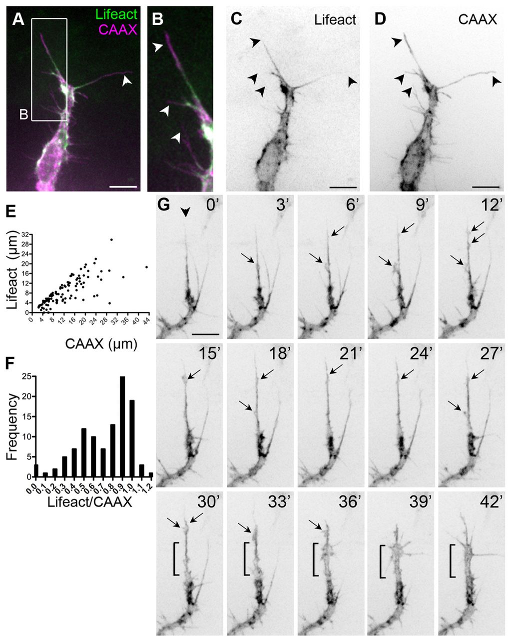
Fig. 3 Dynamic F-actin localisation in filopodia. (A-D) Sprouting front of an ISV from a 30 hpf Tg(Fli1ep:Lifeact-EGFP); Tg(Kdr-l:ras-Cherry)s916 zebrafish embryo. Arrowheads indicate filopodia. (E) Length of the Lifeact signal in a CAAX-labelled filopodium. n=108 filopodia. (F) Frequency distribution of Lifeact/CAAX filopodia length. n=108 filopodia. (G) Dynamics of F-actin in a sprouting ISV from Tg(Fli1ep:Lifeact-EGFP) embryos. Lamellipodia-like structures (arrows) emanate from the most persistent filopodium (arrowhead) and give rise to a rapid expansion in vessel volume (from 30 to 42 minutes, bracket). Scale bars: 10 μm.
Image
Figure Caption
Acknowledgments
This image is the copyrighted work of the attributed author or publisher, and
ZFIN has permission only to display this image to its users.
Additional permissions should be obtained from the applicable author or publisher of the image.
Full text @ Development

