
|
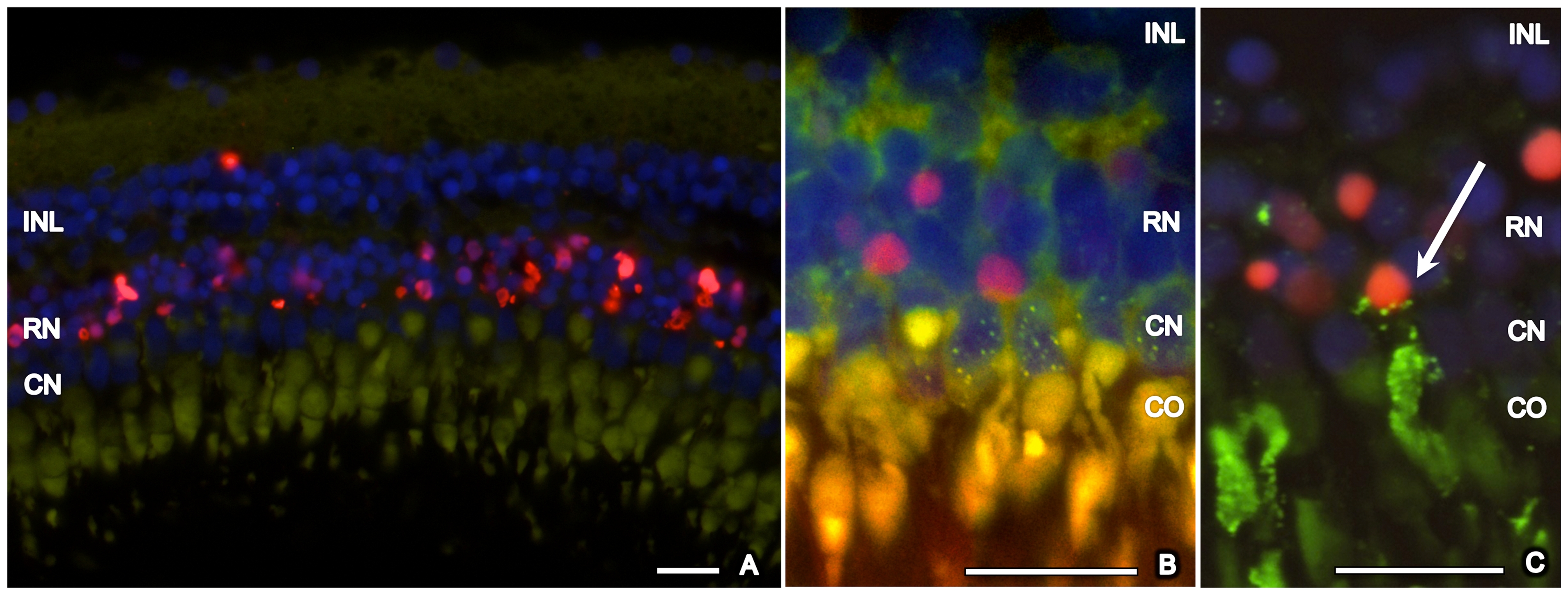
Fig. 3
TUNEL staining of zebrafish retina, 3 days after exposure to MNU 150 mg/l.
A. TUNEL-positive cells (red) are localized in the outer nuclear layer. Strong autofluorescence (green) of a cone outer segment allows identification of the corresponding nearby cone nucleus. Based on this assessment, cone photoreceptors are TUNEL-negative. B. Immunhistochemistry with zpr-1 (staining double cones, green dotted) combined with TUNEL staining (red) confirmed that cone photoreceptors are TUNEL-negative. C. Immunhistochemistry with rhodopsin (staining rods, green) and TUNEL staining (red). Co-Localization of rhodopsin and TUNEL exemplarily shows a dying rod photoreceptor (arrow). Cell nuclei are stained with DAPI (blue). INL (inner nuclear layer), RN (rod nuclei), CN (cone nuclei), CO (cone outer segment). Scale bar indicates 50 μm.