
Fig. S5
- ID
- ZDB-IMAGE-130907-25
- Publication
- Laux et al., 2013 - Circulating Bmp10 acts through endothelial Alk1 to mediate flow-dependent arterial quiescence
- All Figures
- Figures for Laux et al., 2013
|
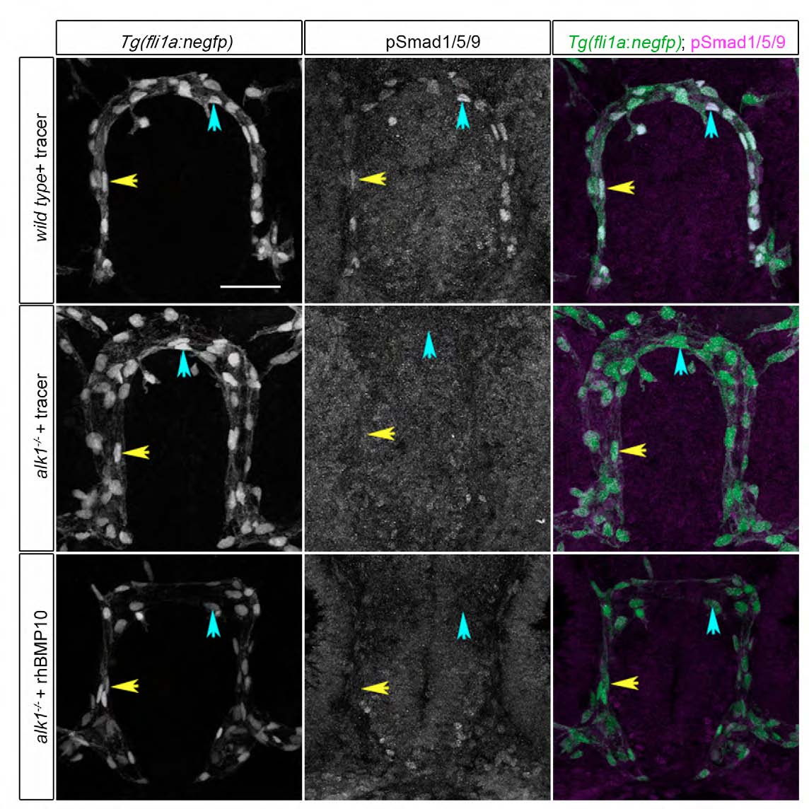
Fig. S5
rhBMP10 cannot induce pSmad1/5/9 in the absence of alk1. Intravascular injections of tracer alone or tracer + 2 nl 10 μM rhBMP10 were performed at 28 hpf into wild-type or alk1 mutant embryos. Images show pSmad1/5/9 expression (middle column) in endothelial cells (nuclei marked by fli1a:nEGFP transgene, left column) at 36 hpf. In merge (right column), EGFP-expressing endothelial cell nuclei are green, pSmad1/5/9 immunofluorescence is magenta. Yellow and blue arrows indicate endothelial cells in the CaDI and BCA, respectively. 2D confocal projections of 50 μm frontal sections, dorsal upwards. Scale bar: 50 μm. See Table S1 for fluorescence quantitation.