
|
Fig. 1
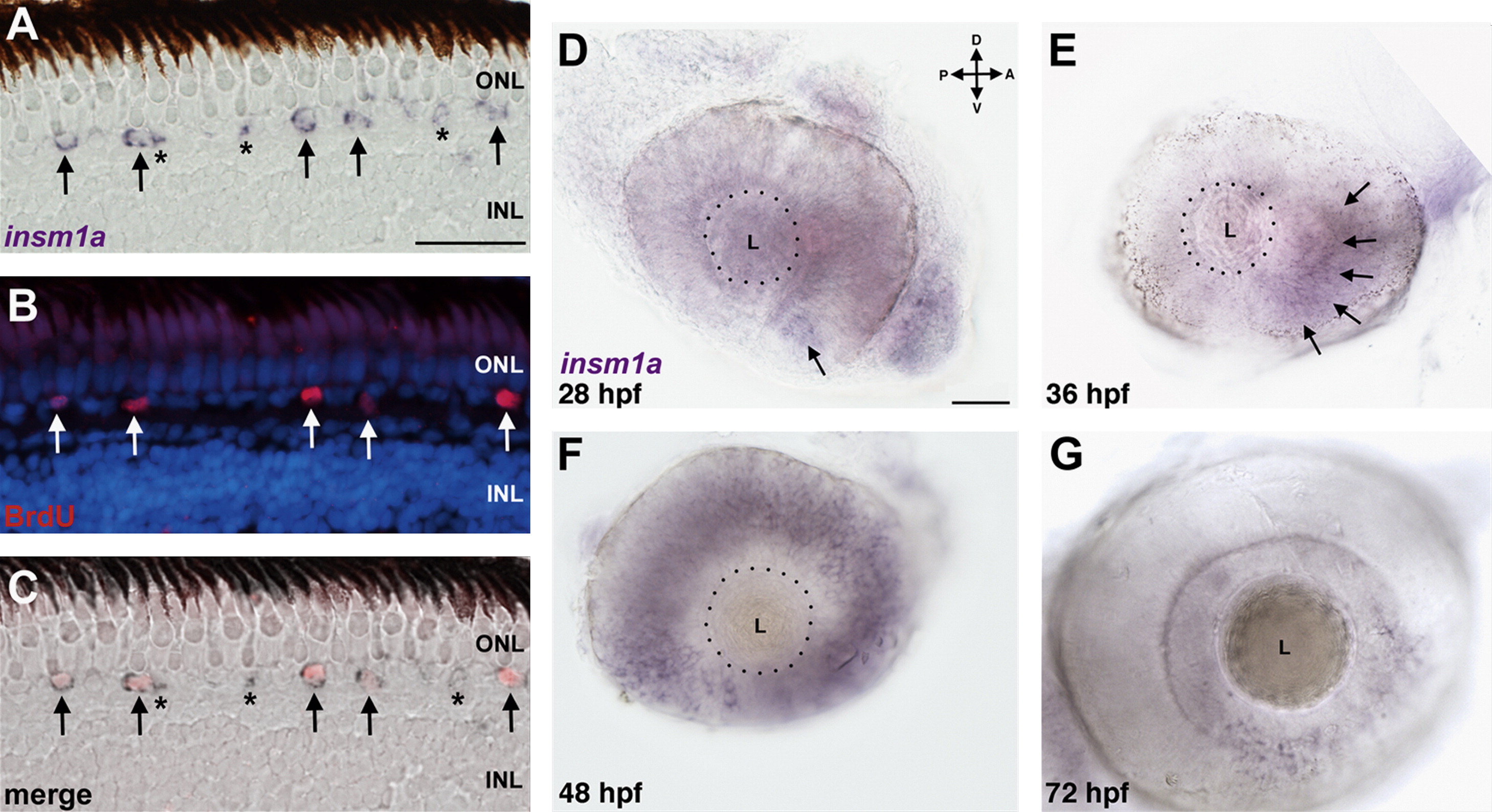
Insm1a is expressed in rod progenitor cells and in the developing retina. (A–C) Expression of insm1a in XOPS-mCFP retinas. (A) In situ hybridization with an antisense probe for insm1a was performed on retinal cryosections of XOPS-mCFP retinas after a 4 h exposure to BrdU. (B) Immunolabeling with anti-BrdU identified rod progenitor cells at the base of the ONL. (C) Overlay of in situ and immunolabeling demonstrates that many, but not all insm1a+ cells are also BrdU+. Arrows indicate insm1a+/BrdU+ cells; asterisks indicate insm1a+/BrdU cells. (D–G) Whole mount in situ hybridization of wild type embryos during development. (D) Insm1a expression was observed in the ventro-nasal patch between 24 and 28 hpf (arrow), (E) and insm1a expression expanded counterclockwise to the dorso-nasal retina at 36 hpf (arrows). (F) By 48 hpf, expression of insm1a had progressed to the dorso-temporal quadrant. (G) At 72 hpf, insm1a expression was only observed adjacent to the proliferative marginal zone at the retinal periphery. (A, scale bar=25 μm; D, scale bar=50 μm; D, dorsal; V, ventral; A, anterior; P, posterior; ONL, outer nuclear layer; INL, inner nuclear layer: L, lens; hpf, hours post fertilization.)
Reprinted from Developmental Biology, 380(2), Forbes-Osborne, M.A., Wilson, S.G., and Morris, A.C., Insulinoma-associated 1a (Insm1a) is required for photoreceptor differentiation in the zebrafish retina, 157-171, Copyright (2013) with permission from Elsevier. Full text @ Dev. Biol.