
Image
|
Figure Caption
Fig. 4
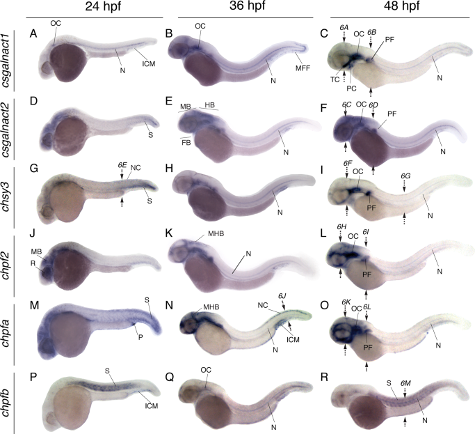
Whole mount in situ hybridization of CS/DS glycosyltransferases at 24–48 hpf during zebrafish development. All images represent lateral views showing the mRNA levels of csgalnact1 (A–C), csgalnact2 (D–F), chsy3 (G–I), chpf2 (J–L), chpfa (M–O), and chpfb (P–R). Arrows indicate the position for sections shown in Figure 6. FB, forebrain; HB, hindbrain; ICM, intermediate cell mass; MB, midbrain; MFF, median fin fold; MHB, midbrain-hindbrain boundary; NC, neural crest; OC, otic capsule; P, proctodeum; PD, pronephric duct; PF, pectoral fin; PC, pharyngeal cartilage; R, retina; S, somites; TC, trabecula cranii.
Figure Data
Acknowledgments
This image is the copyrighted work of the attributed author or publisher, and
ZFIN has permission only to display this image to its users.
Additional permissions should be obtained from the applicable author or publisher of the image.
Full text @ Dev. Dyn.