
Fig. 1
- ID
- ZDB-IMAGE-130808-50
- Genes
- Antibodies
- Publication
- Lee et al., 2013 - An exclusively mesodermal origin of fin mesenchyme demonstrates that zebrafish trunk neural crest does not generate ectomesenchyme
- All Figures
- Figures for Lee et al., 2013
|
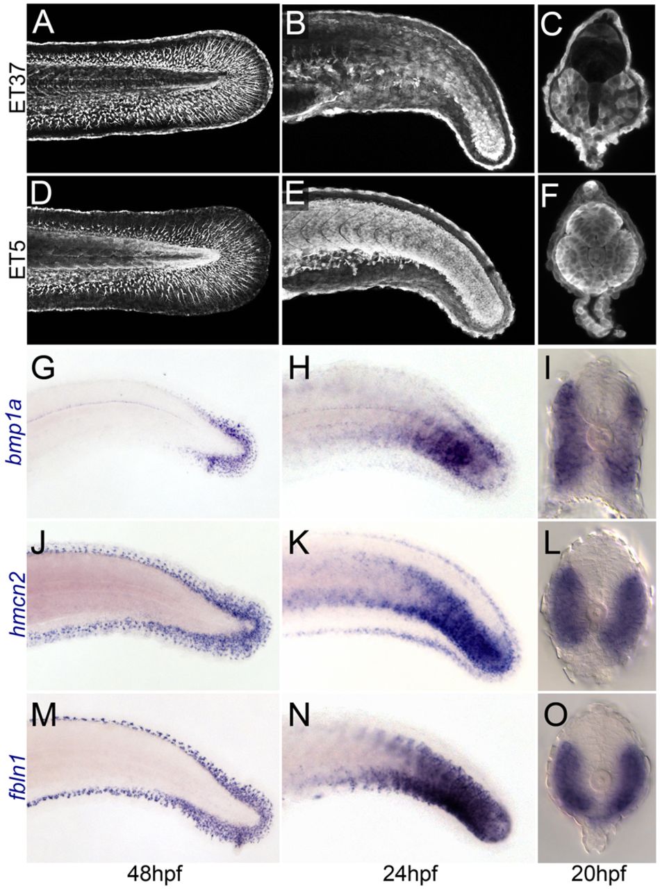
Fig. 1
Paraxial mesoderm expression often precedes fin mesenchyme expression. (A-F) Confocal images of the trunk and tail regions of the ET37 (A-C) and ET5 (D-F) zebrafish lines immunofluorescently stained for eGFP. Lateral views at 48 hpf (A,D) and 24 hpf (B,E) show that expression in fin mesenchyme follows earlier expression in the mesoderm. Transverse confocal images of the trunk of ET37 (C) and ET5 (F) at 20 hpf show expression in paraxial mesoderm as well as at other sites. (G-O) Micrographs of embryos stained by in situ hybridisation with probes against bmp1a (G-I), hmcn2 (J-L) and fbln1 (M-O). Lateral views show the fin mesenchyme expression at 48 hpf (G,J,M) and the preceding mesodermal expression at 24 hpf (H,K,N). Mesodermal expression is paraxial as shown in images of transverse sections of 20-hpf embryos viewed with Nomarski optics (I,L,O).