
IMAGE
Fig. 3
Image
|
Figure Caption
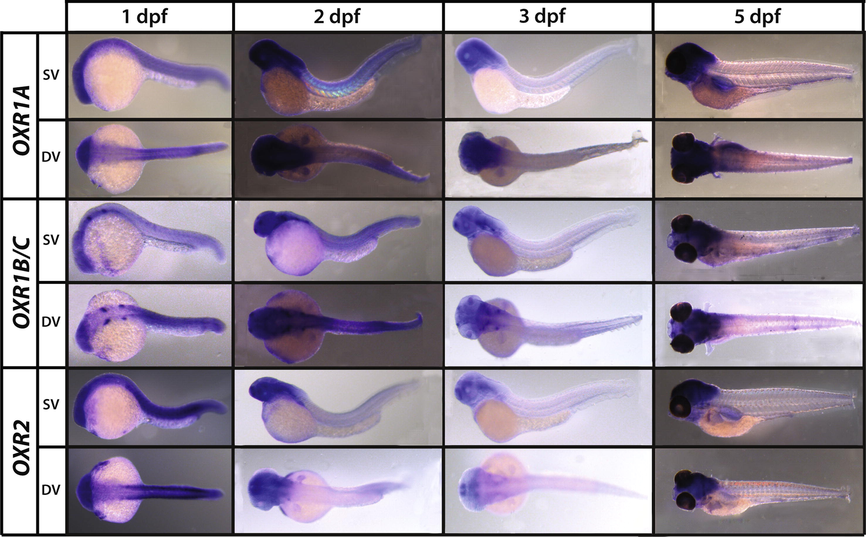
Fig. 3 LysMD (panel A) and OXR (panel B) genes expression patterns during stages of zebrafish development. Whole mount in situ hybrizations using alkaline phosphatase detection and a blue chromogenic substrate. Embryos are orientated with the anterior to the left and dorsal to the top and shown in side view (SV) or dorsal view (DV) at four developmental stages (1,2,3 and 5 dpf).
Figure Data
Acknowledgments
This image is the copyrighted work of the attributed author or publisher, and
ZFIN has permission only to display this image to its users.
Additional permissions should be obtained from the applicable author or publisher of the image.
Reprinted from Gene expression patterns : GEP, 13(7), Laroche, F.J., Tulotta, C., Lamers, G.E., Meijer, A.H., Yang, P., Verbeek, F.J., Blaise, M., Stougaard, J., and Spaink, H.P., The embryonic expression patterns of zebrafish genes encoding LysM-domains, 212-24, Copyright (2013) with permission from Elsevier. Full text @ Gene Expr. Patterns