
Fig. 4
- ID
- ZDB-IMAGE-130726-36
- Genes
- Publication
- Melvin et al., 2013 - A morpholino-based screen to identify novel genes involved in craniofacial morphogenesis
- All Figures
- Figures for Melvin et al., 2013
|
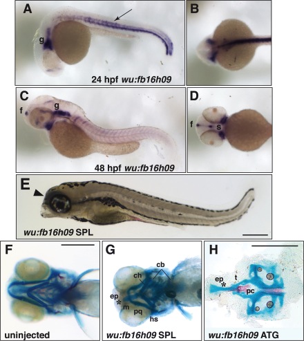
Fig. 4 Neurocranium defects elicited by knock-down of wu:fb16h09. A–D: In situ hybridization for expression of wu:fb16h09. (A) Lateral view and (B) dorsal view of a 24hpf embryo showing expression of wu:fb16h09 in neural tissues of the head (g; ganglia) and trunk hypochord, middle of the somite, and ventral spinal cord (arrow). (C) Lateral view and (D) ventral view of a 48hpf zebrafish showing expression of wu:fb16h09 in the forebrain (f), trigeminal, and other cranial ganglia (g) and regions flanking the presumptive mouth (s, stomodeum). E: Lateral view of a 5dpf larva after injection with 10 ng wu:fb16h09 Morpholino. Arrowhead indicates collapse of the head rostral to the eye. F: Ventral view of the craniofacial skeleton of an uninjected larva at 5dpf. G: Ventral view of 5dpf skeleton of a wu:fb16h09 SPL morphant showing loss of the ethmoid plate and fusion of the trabeculae. (Note that we do not observe cartilage fusions in the viscerocranium with this Morpholino.) H: Flat mount of the neurocranium of wu:fb16h09 ATG morphant after injection with 10 ng ATG Morpholino. Asterisk indicates reduced/absent ethmoid plate and fused trabeculae. Other abbreviations as in Figure 1.