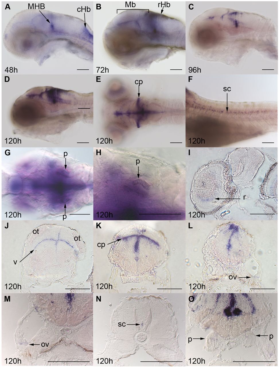
Fig. 2 ZF kcnj10a is expressed in brain, otic vesicles and pronephros. At 24 hpf, kcnj10a expression cannot be detected by in situ hybridisation (not shown) but, by 48 hpf (A), kcnj10a is expressed strongly in the mid-hindbrain boundary (MHB) and caudal hindbrain (cHb), and weakly in the midbrain and rostral hindbrain. (A–F) Until 120 hpf, kcnj10a expression becomes stronger, especially in the midbrain and rostral hindbrain (see Mb and rHb labels in B), and in the cerebellum (cp in E). More posteriorly, expression can be seen in the spinal cord (sc in F), but there is no evidence of expression in the lateral line. (I–O) Transverse sections at 120 hpf demonstrate kcnj10a expression in the inner nuclear layer of the retina (r in I) and reveal the majority of midbrain (J), hindbrain (K,L,M) and spinal cord (N) expression to be adjacent to the ventricles (for example, v in J). kcnj10a is also expressed in the cerebellum (cp in E,K), otic vesicle (ov in L,M) and weakly in the pronephros (p in G,H,O). (A–H) Whole-mount embryos with H being a higher magnification image of the pronephros (p) shown in G. (A–D,F) Lateral views with anterior to the left and dorsal up. (E,G,H) Dorsal views with anterior to the left. (I–L) Progressively more posterior transverse sections. (M–O) Higher magnification images of the otic vesicle, spinal cord and pronephros. ot, optic tectum. Scale bars: 100 μm.
Image
Figure Caption
Figure Data
Acknowledgments
This image is the copyrighted work of the attributed author or publisher, and
ZFIN has permission only to display this image to its users.
Additional permissions should be obtained from the applicable author or publisher of the image.
Full text @ Dis. Model. Mech.

