
Fig. 2
- ID
- ZDB-IMAGE-130709-8
- Genes
- Publication
- Chandrasekar et al., 2013 - The Zebrafish Orthologue of the Dyslexia Candidate Gene DYX1C1 Is Essential for Cilia Growth and Function
- All Figures
- Figures for Chandrasekar et al., 2013
|
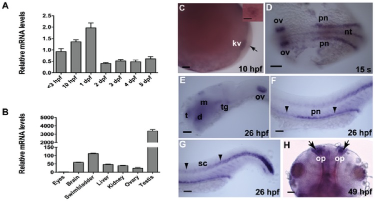
Fig. 2 Expression of dyx1c1 mRNA during embryonic development and in adult tissues.
qPCR analysis of the transcript levels of dyx1c1 during embryonic development (A) and in adult tissues (B). Whole-mount in situ hybridization showed that dyx1c1 is expressed in KV at 10 hpf (C). Inset in C is a close-up view of KV. At 15-somites, dyx1c1 was expressed specifically in the otic vesicle, pronephros and neural tube (D). At 26 hpf dyx1c1 was detected in the brain and is still maintained in otic vesicle, pronephros and spinal canal (E–G). Later at 49 hpf, dyx1c1 was visible in the olfactory placode (H). Panels E–G show lateral views of embryos. Panels D and H show dorsal and ventral views of embryos, respectively. Scale bars indicate 100 μm. Abbreviations: KV, Kupffer’s vesicle: nt, neural tube: pn, pronephros: t, telencephalon: d, diencephalon: m, midbrain: tg, tegmentum: ov, otic vesicle: sc, spinal canal: op, olfactory placode.