
|
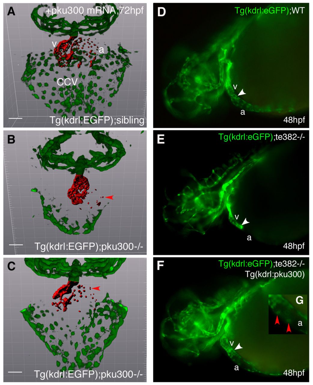
Fig. 4 Both pku300 mRNA and transgenic expression of pku300 in the endocardium rescued the respective mutant phenotypes of scopku300 or scote382. (A-C) The endocardium in the atrium, ventricle and CCV were labeled by Tg(kdrl:eGFP). The endocardium was projected as red pseudocolor with Imaris software. Compared with a wild-type sibling (A), pku300 mRNA rescued the atrial endocardium and CCV of a scopku300 mutant embryo (C). Nine homozygous scopku300 mutants out of 182 offspring from heterozygous scopku300 crosses showed little or no rescue in the atrial endocardium and CCV (B). Wild-type sibling (A) and homozygous scopku300 mutants (B,C) were confirmed by sequencing. Red arrowheads point to the endocardium. (D-G) The endocardium was labeled by Tg(kdrl:eGFP) in the atrium and ventricle of wild-type embryos (D). Note little endocardium in the atrium of scote382 mutant Tg(kdrl:eGFP) embryos (E), which were partially rescued by overexpressing pku300 using Tg(kdrl:pku300) transgene (F). (G) Higher magnification of the atrium of panel F shows the rescued atrial endocardial cells (red arrowheads). The atrium and ventricle are demarcated with white arrowheads (D-F). a, atrium; v, ventricle; WT, wild type. Scale bars: 50μm.