
Fig. 4
- ID
- ZDB-IMAGE-130524-24
- Genes
- Publication
- Roach et al., 2013 - Loss of ascl1a prevents secretory cell differentiation within the zebrafish intestinal epithelium resulting in a loss of distal intestinal motility
- All Figures
- Figures for Roach et al., 2013
|
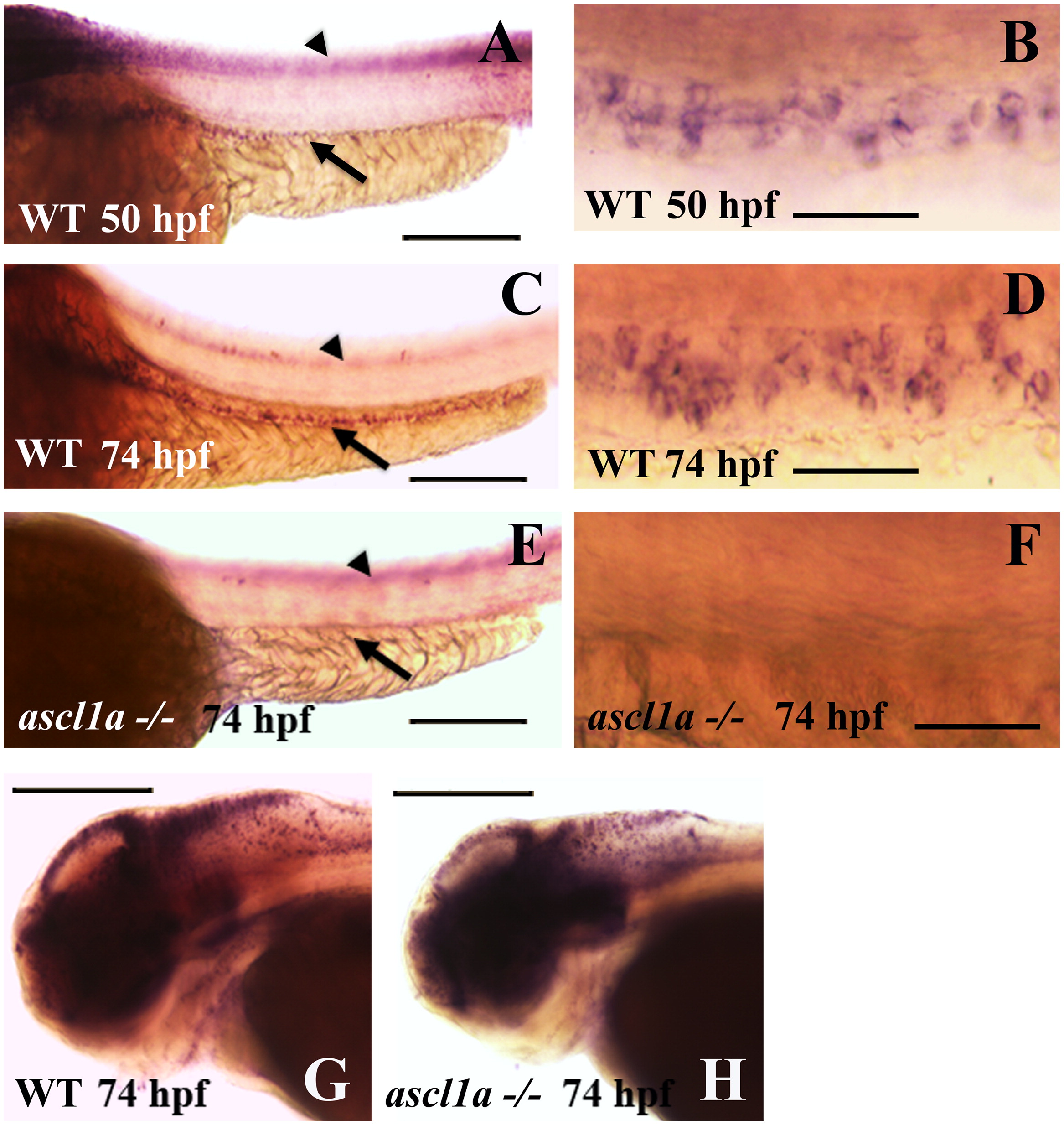
Fig. 4 Intestinal expression of deltaD in WT and ascl1a-/- embryos. deltaD expression in the intestinal epithelium begins on the second day of embryogenesis by 50 hpf ((A), arrow: intestinal expression; arrowhead: neural tube expression out of focal plane) with stronger expression in the proximal intestine. Magnified view of 50 hpf deltaD expression (B) demonstrates small clusters and some isolated intestinal epithelial cells. deltaD expression becomes stronger and more even throughout the intestinal epithelium at 74 hpf ((C): arrow; arrowhead: neural tube expression out of focal plane). Magnified view of 74 hpf (D) demonstrates that deltaD is now expressed in larger clusters and higher numbers of intestinal epithelial cells. ascl1a-/- embryos lack expression of deltaD within the intestinal epithelium at 74 hpf (arrow in (E)) but retain neural tube expression (arrowhead in (E) out of focal plane). Magnified view of ascl1a-/- (F) shows absence of deltaD expression in intestinal epithelium. Within the same ascl1a-/- embryos, deltaD expression in the brain is altered but remains strong (compare WT in (G) to ascl1a-/- in (H)). Anterior is to the left and posterior to the right in all panels. Scale bars: (A), (C), (E), (G), and (H) 200 μm; (B), (D), and (F) 50 μm.
Reprinted from Developmental Biology, 376(2), Roach, G., Heath Wallace, R., Cameron, A., Emrah Ozel, R., Hongay, C.F., Baral, R., Andreescu, S., and Wallace, K.N., Loss of ascl1a prevents secretory cell differentiation within the zebrafish intestinal epithelium resulting in a loss of distal intestinal motility, 171-186, Copyright (2013) with permission from Elsevier. Full text @ Dev. Biol.