
Fig. 5
- ID
- ZDB-IMAGE-130524-20
- Genes
- Antibodies
- Publication
- Roach et al., 2013 - Loss of ascl1a prevents secretory cell differentiation within the zebrafish intestinal epithelium resulting in a loss of distal intestinal motility
- All Figures
- Figures for Roach et al., 2013
|
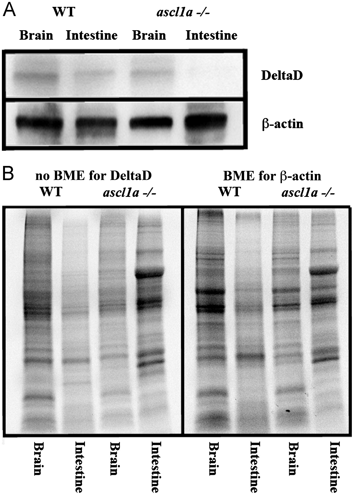
Fig. 5 Intestinal expression of DeltaD in WT and ascl1a-/- embryos. (A) Both mutant and WT 5 dpf intestines and brains were dissected for western blot. Brains for both WT and ascl1a-/- demonstrate similar levels of DeltaD and the loading control β-actin. ascl1a-/- intestines lack DeltaD while there are similar levels of β-actin compared to WT. DeltaD is approximately 100 kD while β-actin is 55 kD. (B) Total protein as visualized by induced fluorescence on stain-free gel. Proteins are visualized after electrophoresis but before transfer to membrane as an alternative loading control to β-actin. The protein gel for DeltaD was run without β-mecaptoethanol (BME) and shown to the left while protein gel for β-actin was run with β-mecaptoethanol (BME) and is shown to the right. The same volume of protein from the same sample was loaded on both the BME and no BME gels.
Reprinted from Developmental Biology, 376(2), Roach, G., Heath Wallace, R., Cameron, A., Emrah Ozel, R., Hongay, C.F., Baral, R., Andreescu, S., and Wallace, K.N., Loss of ascl1a prevents secretory cell differentiation within the zebrafish intestinal epithelium resulting in a loss of distal intestinal motility, 171-186, Copyright (2013) with permission from Elsevier. Full text @ Dev. Biol.