
|
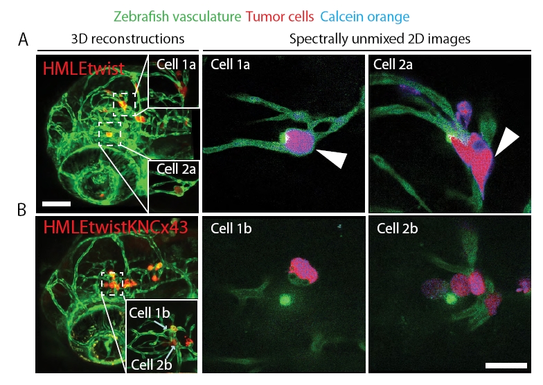
Fig. S5
HMLEtwist cells exchange calcein orange dye with brain endothelial cells in connexin 43-dependent manner in vivo. (A) HMLEtwist or (B) HMLEtwistKNcx43 cells were preloaded with calcein orange dye (shown as blue) and injected into the circulation of zebrafish and allowed to form microtumors along the vasculature (green). Dye transfer to the endothelium was monitored in live animals using confocal microscopy. Left panels show 3D reconstructions (20x) of the zebrafish head region with seeded tumor cells. Right panels show higher magnification (60x) of tumor cells delineated with dashed squares. Note the strong presence of the calcein orange dye within the endothelial cells (white arrows) that are located next to HMLEtwist cells, but not in the endothelium adjacent to HMLEtwistKNcx43 cells. Bars = 100 μm in A and 20 μm in B.