
Fig. 3
- ID
- ZDB-IMAGE-130502-14
- Antibodies
- Publication
- Song et al., 2013 - Pou5f1-dependent EGF expression controls e-cadherin endocytosis, cell adhesion, and zebrafish epiboly movements
- All Figures
- Figures for Song et al., 2013
|
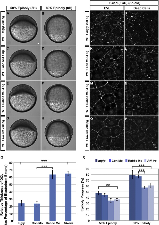
Fig. 3 Inhibition of Rab5c Affects Epiboly(A–H) Live WT embryos injected with mRNA encoding membrane-tagged GFP (mGFP) (A and B), control morpholino (con Mo) (C and D), Rab5c MO (E and F), or RN-tre mRNA (G and H) are shown at stages corresponding to WT 50% epiboly and 80% epiboly. Lateral views, animal pole to the top. Scale bar, 50 μm.(I–P) E-cad subcellular localization in mgfp (I and J), control Mo (K and L), Rab5c Mo (M and N), or RN-tre mRNA (O and P) injected WT embryos at shield stage (confocal image of anti-E-cad whole mount immunofluorescence). Animal views. Scale bar, 10 μm.(Q and R) Quantification of (Q) relative DCL thickness at 50% epiboly and (R) epiboly progress at 50% epiboly and 80% epiboly stages (Error bars show SEM; p < 0.01, p < 0.001; n = 12 embryos each).See also Figure S3.
Reprinted from Developmental Cell, 24(5), Song, S., Eckerle, S., Onichtchouk, D., Marrs, J.A., Nitschke, R., and Driever, W., Pou5f1-dependent EGF expression controls e-cadherin endocytosis, cell adhesion, and zebrafish epiboly movements, 486-501, Copyright (2013) with permission from Elsevier. Full text @ Dev. Cell