
IMAGE
Fig. S2
- ID
- ZDB-IMAGE-130430-2
- Publication
- Choudhry et al., 2013 - DiGeorge Syndrome Gene tbx1 Functions through wnt11r to Regulate Heart Looping and Differentiation
- All Figures
- Figures for Choudhry et al., 2013
Image
|
Figure Caption
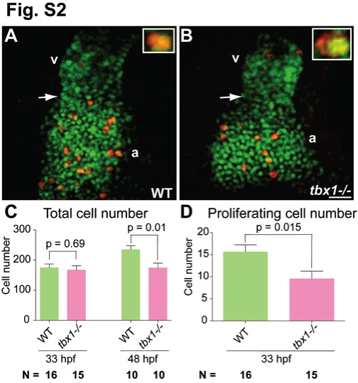
Fig. S2 Proliferation defects in tbx1-/- mutants. (A, B) Confocal projections of hearts from cmlc2:gfp (all heart cells are green) embryos at 33 hpf stained with phospho-histone 3 antibody (proliferating cells are red). The insets in A and B show a magnified view of a co-labeled cell (yellow). (C, D) Plots showing the total (C) and proliferating (D) number of cells in WT and tbx1-/- mutants. N is the total number of embryos. Arrows point to the AVC; v, ventricle; a, atrium. Scale bar: 25 μm.
Figure Data
Acknowledgments
This image is the copyrighted work of the attributed author or publisher, and
ZFIN has permission only to display this image to its users.
Additional permissions should be obtained from the applicable author or publisher of the image.
Full text @ PLoS One