
Fig. 6
- ID
- ZDB-IMAGE-130425-34
- Publication
- Li et al., 2013 - Analysis of a gene regulatory cascade mediating circadian rhythm in zebrafish
- All Figures
- Figures for Li et al., 2013
|
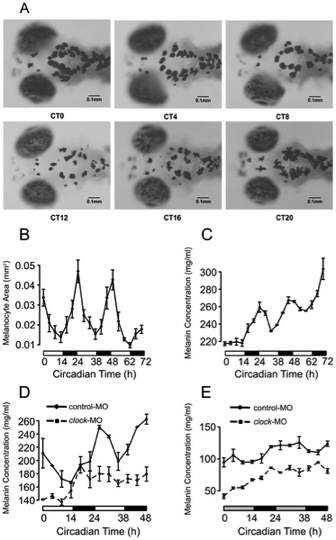
Fig. 6 The circadian rhythm of melanogenesis in larval zebrafish.
(A) Images of 5 dpf WT larval melanocytes in 4 hour intervals over 24 hours under LD conditions. (B) The area of melanocytes in WT larvae showed robust circadian rhythm in LD starting at 4 dpf (p<0.001, Fisher′s g test). (C) Melanin concentrations of WT larvae showed robust circadian rhythm in LD while increasing with time (p<0.002, Fisher′s g test after detrend). (D) The rhythm of melanin concentration was abolished in clock morphants in LD conditions, while the rhythm in control morphants remained robust. (E) Under DD conditions, the circadian rhythms of melanin concentrations in clock morphants and control morphants were not significant.