
|
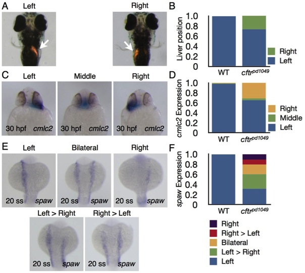
Fig. 2 Organ laterality is disrupted in cftr1049 mutant embryos. Heterozygous cftrpd1049 fish were mated to assess organ laterality. (A) Ventral view of 4 days post-fertilization (dpf) WT and cftrpd1049 mutant larvae expressing dsRed in the liver (arrows). (B) Quantification of liver orientation in WT and cftrpd1049 mutants. Liver orientation is reversed in 27% of homozygous mutants. WT, n=103; cftrpd1049, n=30. (C) Ventral view of representative cftrpd1049 mutants showing cmlc2 expression pattern. (D) Quantification of heart looping in WT and cftrpd1049 mutants. WT, n=408; cftrpd1049, n=138. (E) Dorsal view of cftrpd1049 mutant embryos displaying left, right, left>right, right>left and left=right spaw expression patterns. (F) Quantification of spaw expression in WT and cftrpd1049 mutants. WT, n=146; cftrpd1049, n=73.