
Fig. 3
- ID
- ZDB-IMAGE-130318-31
- Antibodies
- Publication
- Qian et al., 2013 - ENC1-like Integrates the Retinoic Acid/FGF Signaling Pathways to Modulate Ciliogenesis of Kupffer's Vesicle during Zebrafish Embryonic Development
- All Figures
- Figures for Qian et al., 2013
|
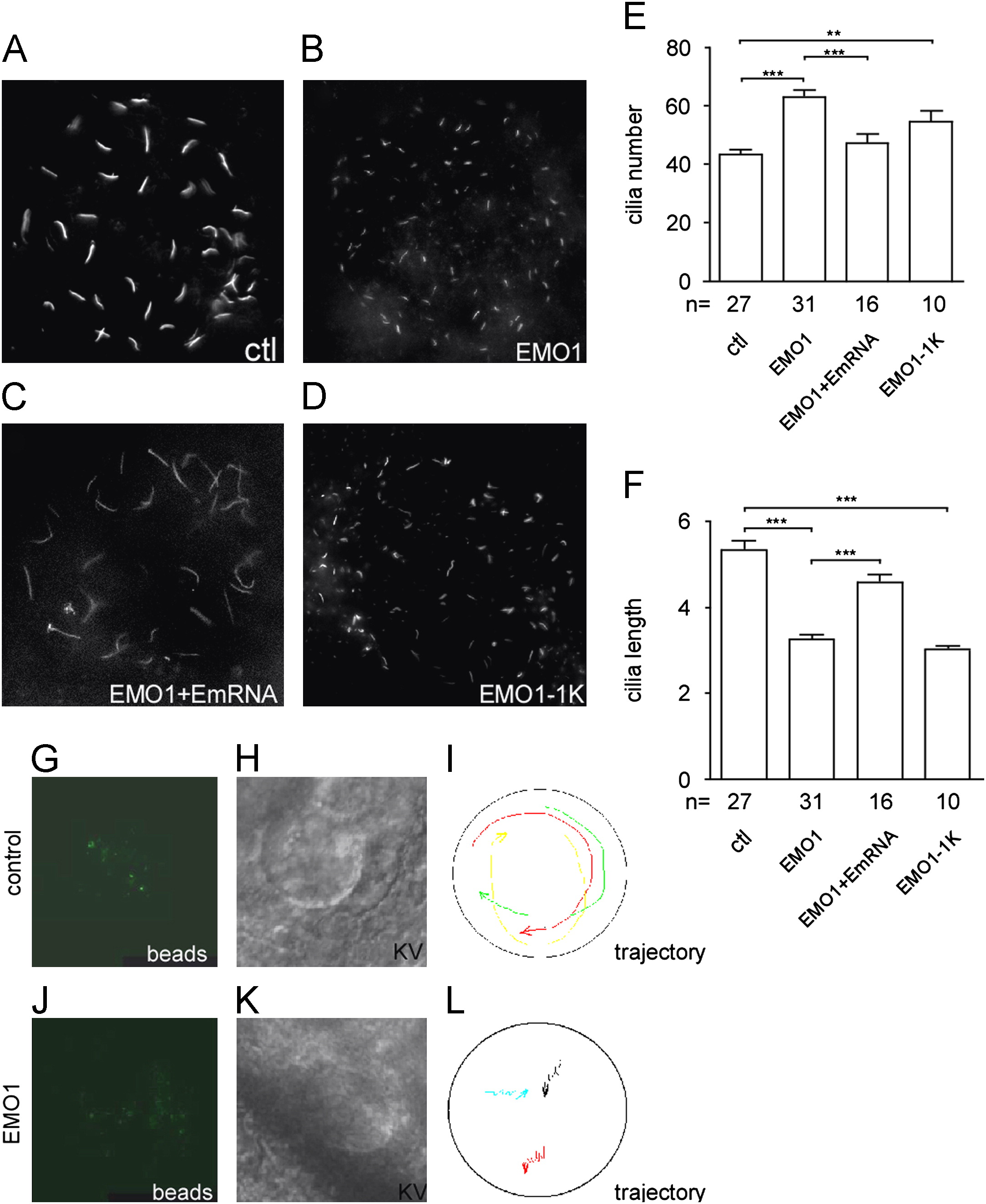
Fig. 3 Enc1l is essential for KV ciliogenesis and KV function. (A–F) Anti-acetylated tubulin antibody is used to visualize the cilia in KV. (A) Control embryos, (B) enc1l-MO1 is injected at 1-cell stage, (C) enc1l-MO1 and enc1l-mRNA are co-injected at 1-cell stage. (D) enc1l-MO1 is injected at the 1000-cell stage (EMO1-1K). (E, F) Statistical graphs show the decrease in cilium length and increase in cilium number. (G–L): Enc1l is required for counterclockwise nodal fluid flow. (G–I) Still images from control-MO embryos and (J–L) enc1l-MO injected embryos. These images are taken from supplement Movie1 and 2, respectively. (G, J): Fluorescent beads in the KV. (H, K): The KV structure from the DIC image. (I, L): Trajectory of the fluorescent beads in KV. **0.005<π<0.01; *** :p<0.005.
Reprinted from Developmental Biology, 374(1), Qian, M., Yao, S., Jing, L., He, J., Xiao, C., Zhang, T., Meng, W., Zhu, H., Xu, H., and Mo, X., ENC1-like Integrates the Retinoic Acid/FGF Signaling Pathways to Modulate Ciliogenesis of Kupffer's Vesicle during Zebrafish Embryonic Development, 85-95, Copyright (2013) with permission from Elsevier. Full text @ Dev. Biol.