
|
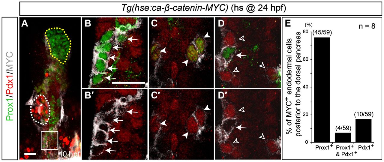
Fig. 4 Wnt/β-catenin signaling cell-autonomously induces hepatoblasts.
Tg(hse:ca-β-catenin-MYC) embryos were heat-shocked at 24hpf, harvested at 40hpf, and processed for anti-Prox1 (green), anti-Pdx1 (red) and anti-MYC (gray) staining. ca-β-catenin-expressing cells were detected by anti-MYC antibodies. (A,B) Most MYC+ cells in the endoderm posterior to the dorsal pancreas expressed Prox1 (B, arrows) but not Pdx1. Since the Prox1 antibody has a background staining problem, cytoplasmic or membrane-like staining is background staining, whereas nuclear Prox1 staining is real staining. White and yellow dotted lines in A outline the dorsal pancreas and liver, respectively. Higher magnification images of the square region in A are shown in B,B2. (C,C2) Some MYC+ cells expressed both Prox1 and Pdx1 (arrowheads). (D,D2) Weak MYC+ cells were Prox1/Pdx1+, whereas strong MYC+ cells were Prox1+/Pdx1 (open arrowheads versus arrow). Ventral views, anterior up. Scale bars, 20µm. (E) Quantification of MYC+ cells in the endoderm posterior to the dorsal pancreas based on Prox1 and Pdx1 expression. n indicates the number of embryos.