
|
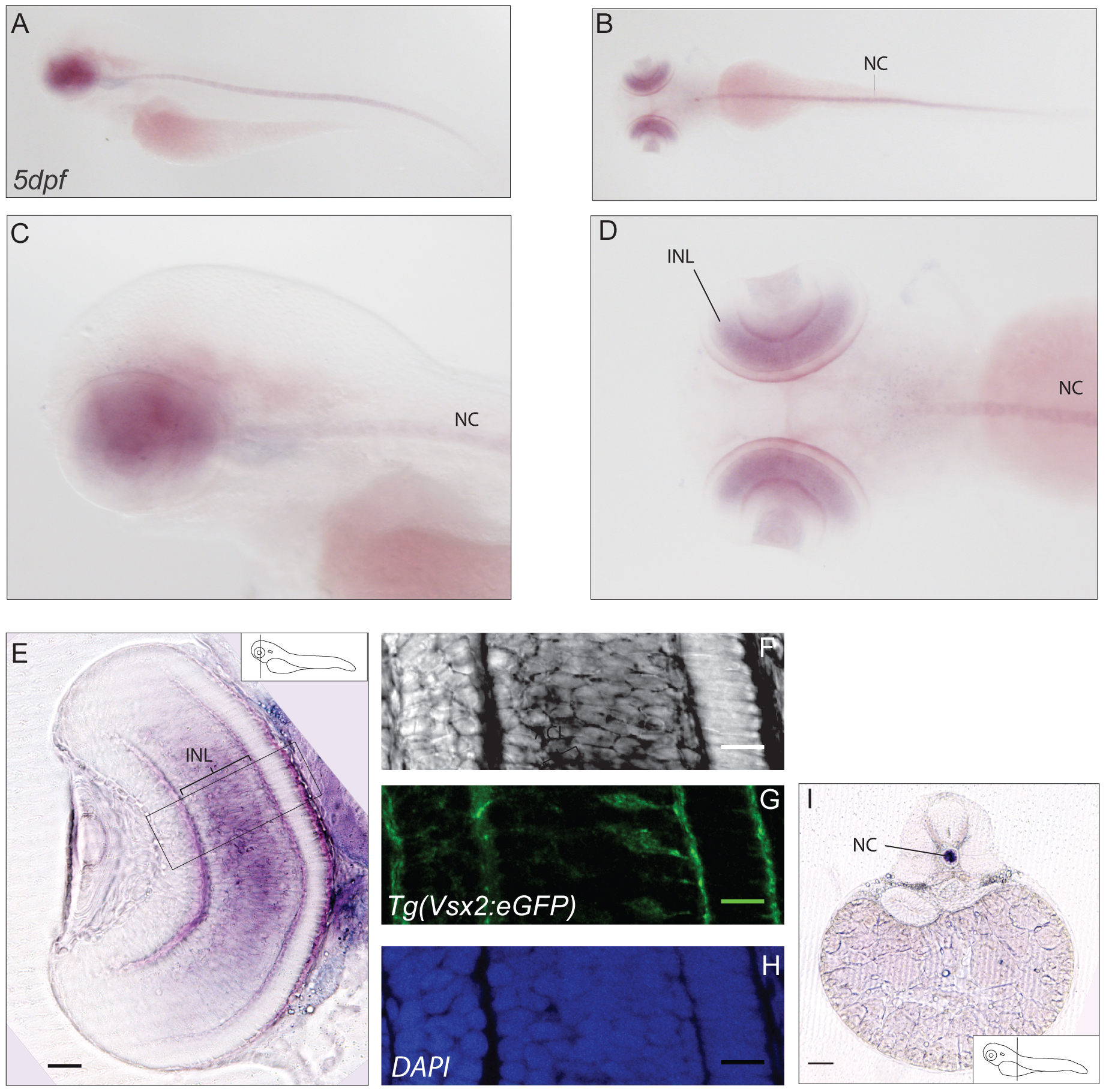
Fig. 4 Expression pattern of cabp2a.
(A–D) In situ hybridisation showing expression of cabp2a in 5dpf larvae. Lateral views (A,C) , dorsal views (B, D). Cabp2a antisense probe exhibits diffuse retinal localisation; a strong staining is also present along the notochord. (E–H) Cross-section of a retina in the Tg(Vsx2:GFP) transgenic line. (E) Epifluorescence image of a sectioned retina, showing prominent expression of cabp2a in the bipolar cell layer. Scale bar: 50 µm. (F–H) Confocal images of the area selected in E. (F) In situ signal in bright field, (G) GFP signal localized in bipolar cells, (H) DAPI. Scale bar: 20 µm (I) Cross-section exhibiting in situ staining in the notochord. Scale bar: 100 µm. INL: inner nuclear layer, NC: notochord.