
Fig. 5
- ID
- ZDB-IMAGE-130123-52
- Genes
- Antibodies
- Publication
- Schweitzer et al., 2013 - Sim1a and Arnt2 contribute to hypothalamo-spinal axon guidance by regulating Robo2 activity via a Robo3-dependent mechanism
- All Figures
- Figures for Schweitzer et al., 2013
|
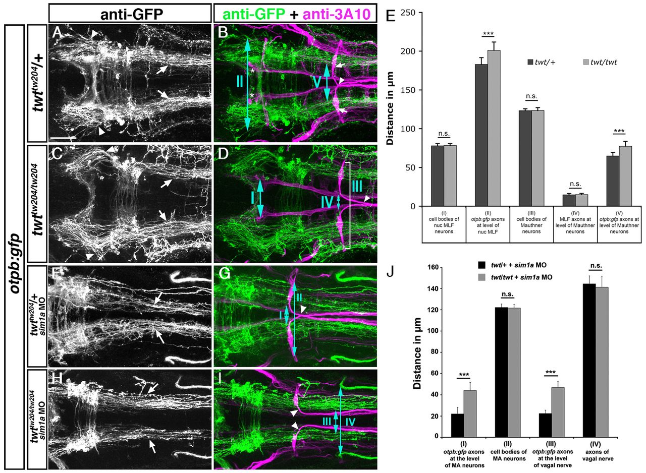
Fig. 5 Robo3 is required for lateral positioning of otpb:gfp-positive longitudinal axons. Dorsal views of confocal hindbrain z-projections (72 hpf) are shown. Anterior is towards the left. (A,B) In twt/+ embryos, otpb:gfp longitudinal axons in the midbrain (A, arrowheads) and hindbrain (A, arrows) grow by a defined distance towards the midline. Arrows in B indicate Mauthner neuron somata. (C,D) In twt/twt embryos, otpb:gfp longitudinal axons project more laterally in the midbrain (C, arrowheads) and hindbrain (C, arrows). (E) Quantification of the separation between otpb:gfp longitudinal axons in mid- and hindbrain (double-headed arrows, II and V in B) and between nucMLF somata (I, see asterisks in B), MLF axons (IV) or MA neurons (III) in twt/+ or twt/twt embryos. (F,G) In sim1a MO-injected twt/+ embryos, otpb:gfp longitudinal axons are medially displaced (F, arrows). (H,I) In sim1a MO-injected twt/twt embryos, otpb:gfp longitudinal axons project more laterally (H, arrows). (J) Quantification of the distance between MA neurons (II, marked in G) and vagal axons (IV in H), or of the distance between otpb:gfp longitudinal axons (I in H and III in G) in sim1a MO-injected twt/+ and twt/twt embryos. Arrowheads in B,D,G,I indicate pathfinding of Mauthner neurons in twt/+ and twt/twt embryos. *P<0.001. Scale bar: 50 μm.