
Fig. 3
- ID
- ZDB-IMAGE-130109-8
- Publication
- Ichimura et al., 2012 - Structural disorganization of pronephric glomerulus in zebrafish mpp5a/nagie oko mutant
- All Figures
- Figures for Ichimura et al., 2012
|
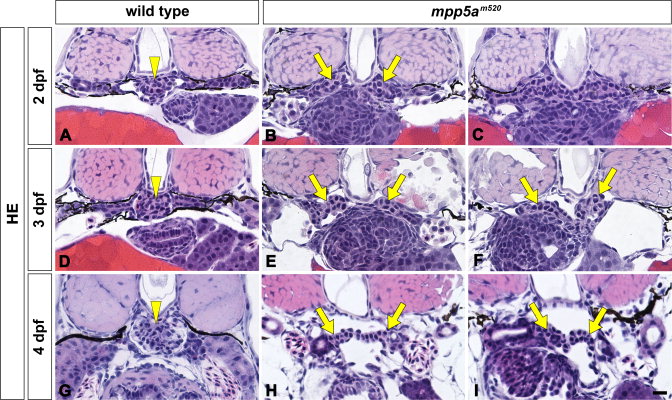
Fig. 3 mpp5am520 mutant displays structural disorganization of pronephric glomerulus. Pronephric glomerular structure is shown by hematoxylin-eosin stained JB-4 section at 2 dpf (A-C), 3 dpf (D–F), and 4 dpf (G-I). In wild type siblings, a pair of glomerular primordia has merged to form a single glomerulus (arrowheads) beneath the notochord at 2 dpf (A), 3 dpf (D), and 4 dpf (G). In 2-dpf mpp5am520 mutants, a pair of glomerular primordia retains epithelial vesicular structure that does not merge beneath the notochord (arrows in B, C). At 3 and 4 dpf mpp5am520 mutants still exhibited two separated podocyte masses (arrows in E, F, H, I). Scale bar = 10 μm.