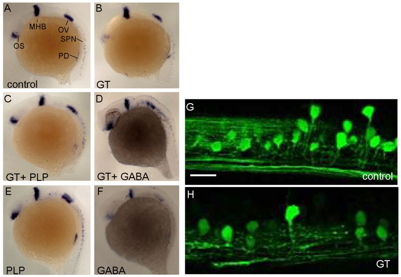
Fig. 6 Ginkgotoxin-induced malformation of optic stalk and spinal cord neuron was partially reversed by PLP or GABA supplementation. Zebrafish embryos were raised in fish water (control) or water containing 500 μM ginkgotoxin added at 6 hpf (GT). (A,B) The development of embryos were characterized at 24 hpf using in-situ hybridization with pax2.1 probe (lateral view with anterior to the left). Development of ostic stalk and spinal cord neuron were interfered with in the ginkgotoxin-treated group (B) and the effect was reversed in the presence of 0.5 mM PLP (C) or 0.5 mM GABA (D). Adding PLP alone did not cause observable changes in embryos (E), whereas adding GABA alone to the water impeded the development of pax2.1-expressing tissues, especially optic stalk, otic vesicle and spinal cord neuron (F). (G,H) Confocal microscopy images of the nortochord in the trunk of Tg(alx:GFP) transgenic embryos at 3 dpf. OS, optic stalk; MHB, midbrain-hindbrain boundary; OV, otic vesicle; SPN, spinal cord neurons; PD, pronephric duct. Scale bar: 8 μm.
Image
Figure Caption
Figure Data
Acknowledgments
This image is the copyrighted work of the attributed author or publisher, and
ZFIN has permission only to display this image to its users.
Additional permissions should be obtained from the applicable author or publisher of the image.
Full text @ Dis. Model. Mech.

