
|
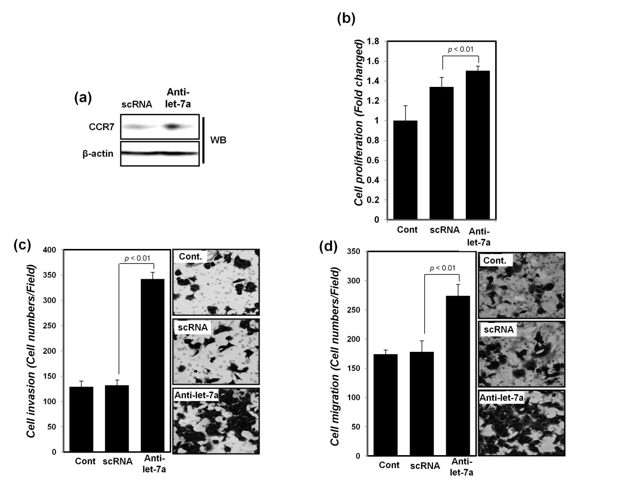
Fig. 4
Cell proliferation, cell migration, and invasion of MCF-7 breast cancer cell lines were increased after transfection with anti-let7a. (a) Protein expression of CCR7 was detected with Western blot analysis after transfection with scRNA and anti-let-7a, respectively. β-actin was used as a normalization control. After transfection with scRNA or anti-let7a, (b) cell proliferation by MTT analysis, (c) cell-migration assays, and (d) invasion assays were performed, as described in Material and Methods, and the results of (c) and (d) presented as a histogram (left panel) with cell photos (right panel). All experiments were performed in triplicate independently.