
|
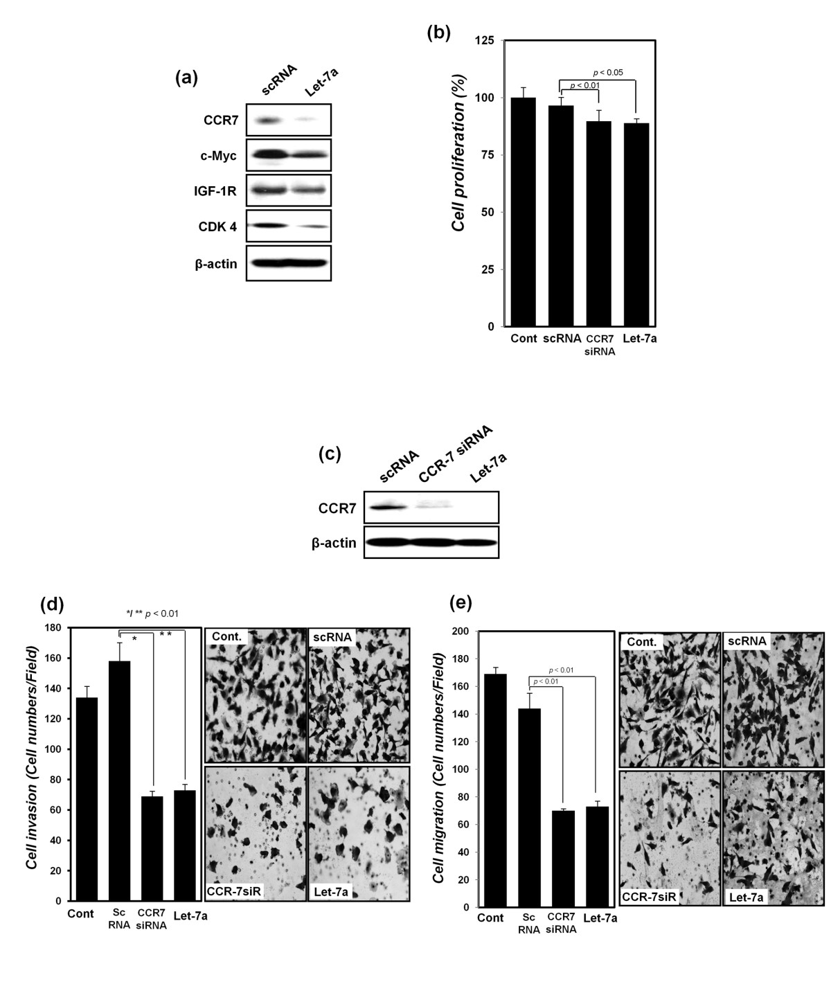
Fig. 3
Detection of changes in the expression of let-7a predicted target proteins and cell proliferation, cell migration, and invasion of MDA-MB-231 breast cancer cells after transfection with synthetic let-7a. (a) After transfection with scRNA or synthetic let-7a, protein expression of CCR7, IGF-1R, c-Myc, and CDK-4 was detected with Western blot analysis. (b) Proliferation of MDA-MB-231 cells was assessed with MTT assay after transfection with scRNA, CCR7 siRNA, and synthetic let-7a, respectively. (c) Protein expression of CCR7 was detected with Western blot analysis after transfection with scRNA, CCR7 siRNA, and synthetic let-7a, respectively. β-actin was used as a normalization control. (d) Cell-migration assays and (e) invasion assays performed after transfection with scRNA, CCR7 siRNA, and synthetic let-7a, as described in Materials and Methods, presented as a histogram (left panel) with cell photos (right panel) All the experiments were performed in triplicate independently.