
Image
|
Figure Caption
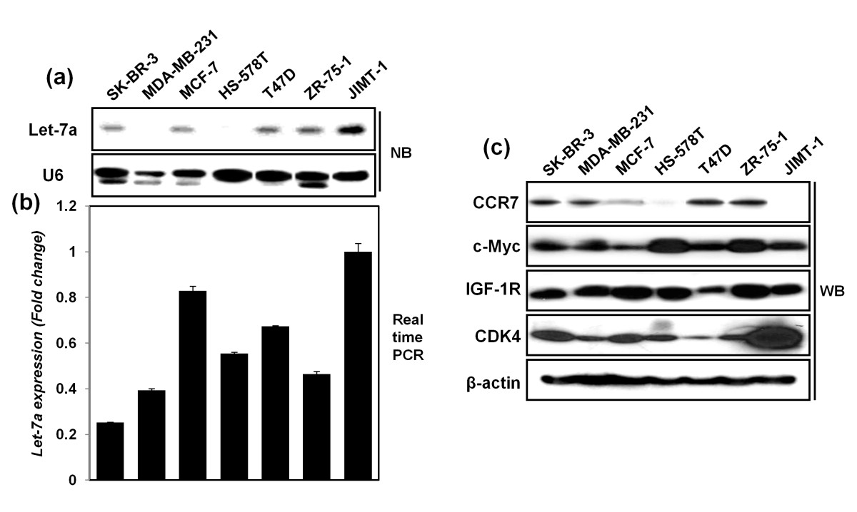
Fig. 2
Detection of basal expression levels of CCR7, IGF-1R, c-Myc, CDK-4, and let-7a in seven breast cancer cell lines. (a-b) Let-7a expression levels in breast cancer cells, detected with Northern blot analysis (NB)(a) and real-time RT-PCR(b). U6 and RNU6B were used as a normalization control. (c) Detection of protein expression levels of CCR7, IGF-1R, c-Myc, and CDK-4 by using Western blot analysis. β-actin was used as a normalization control.
Acknowledgments
This image is the copyrighted work of the attributed author or publisher, and
ZFIN has permission only to display this image to its users.
Additional permissions should be obtained from the applicable author or publisher of the image.
Full text @ Breast Cancer Res.