
Fig. S6
- ID
- ZDB-IMAGE-121011-16
- Publication
- Seiler et al., 2012 - Smooth muscle tension induces invasive remodeling of the zebrafish intestine
- All Figures
- Figures for Seiler et al., 2012
|
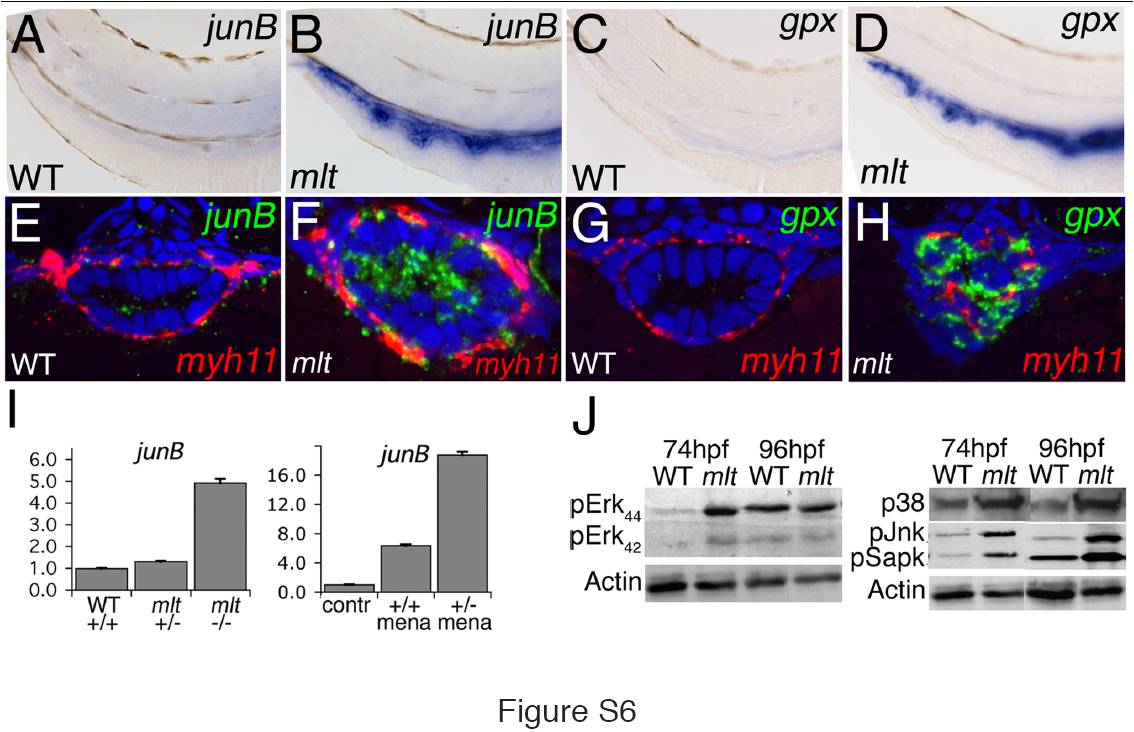
Fig. S6 AP-1 transcription factors, ROS responsive genes, and MAP-Kinase signaling are activated in the mlt intestine. (A–D) Whole mount in situ hybridization shows strong expression (blue) of the AP-1 gene junB and the ROS activated gene gluthatione peroxidase (gpx) in the intestine of 74 hpf mlt (B, D) but not WT (A, C) larvae. n = 15 mlt and wild type larvae examined. (E, F) Histological cross-sections of whole mount specimens processed for fluorescent RNA in situ hybridization show strong junB expression (green) in mlt intestinal epithelial cells with only low level expression in smooth muscle cells (labeled red by myh11 expression). (G, H) Similarly, gpx expression (green) can only be detected in the mlt intestinal epithelium. n = 12 mlt and 12 wild type larvae examined. (I) Quantitative RT-PCR shows increased junB expression in the intestine of mlt homozygotes. junB expression is also increased in menadione treated wild type larvae (+/+), and to a greater degree in mlt heterozygous larvae (versus untreated wild type; contr). (J) Western blot showing phosphorylation of several components of the Map-Kinase signaling pathway in intestines dissected from mlt larvae at 74 hpf and 96 hpf. ERK is prematurely activated at 74 hpf in mlt. p38 (Mapk), Jnk, and Sapk are strongly activated in mlt but not WT larvae at 74 hpf and 96 hpf. beta-Actin, loading control. Western blots are representative of between two and four independent experiments.