
IMAGE
Fig. S1
- ID
- ZDB-IMAGE-121011-11
- Publication
- Seiler et al., 2012 - Smooth muscle tension induces invasive remodeling of the zebrafish intestine
- All Figures
- Figures for Seiler et al., 2012
Image
|
Figure Caption
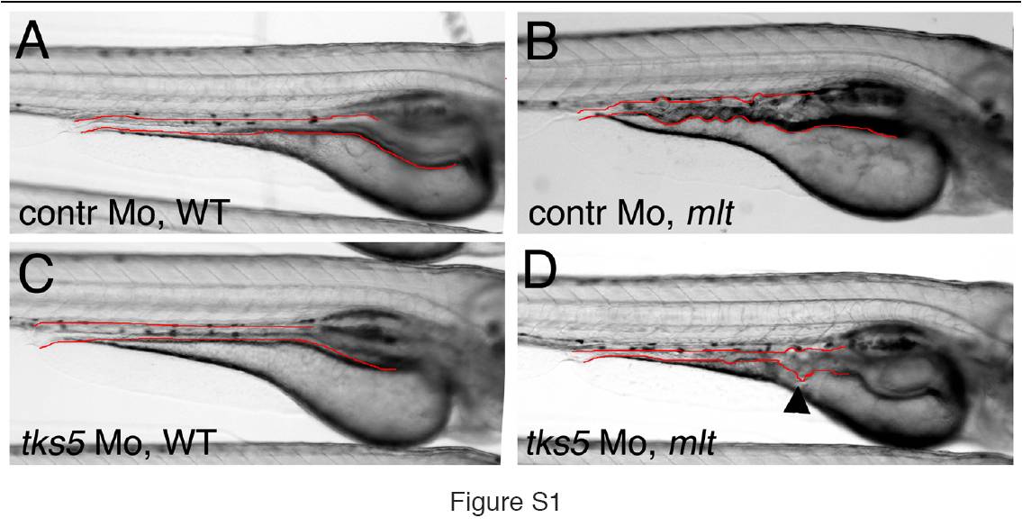
Fig. S1 tks5 knockdown rescues epithelial invasion in the intestine of mlt mutant larvae. (A–C) Lateral images of live 5 dpf larvae. Control morpholino injected mlt larvae show cystic expansion of the intestinal epithelium (intestinal epithelium outlined in red; lateral view) (B), while only a small number of cysts can still be detected in the tks5 morpholino injected mlt larvae (D, arrowhead). The majority of the posterior intestine in these larvae resembles WT (A, C). Findings confirmed histologically (not shown; n = 6 mlt larvae).
Acknowledgments
This image is the copyrighted work of the attributed author or publisher, and
ZFIN has permission only to display this image to its users.
Additional permissions should be obtained from the applicable author or publisher of the image.
Full text @ PLoS Biol.