
Fig. S16
- ID
- ZDB-IMAGE-120824-6
- Publication
- Chen et al., 2012 - Haemodynamics-driven developmental pruning of brain vasculature in zebrafish
- All Figures
- Figures for Chen et al., 2012
|
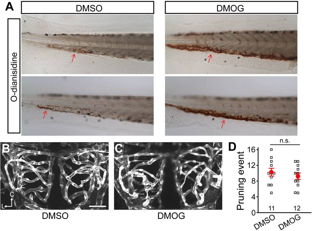
Fig. S16 Effects of DMOG treatment on vessel pruning. (A) o-Dianisidine staining showing that DMOG treatment increases the amount of blood cells in treated embryos. DMSO (0.2%) or DMOG (0.2 mM) was bath-applied during 2–3 dpf. (B and C) Effect of DMOG treatment on vessel pruning of larval zebrafish midbrain. Projected confocal images showing midbrain vasculature of DMSO- (B) and DMOG-treated (C) zebrafish larvae at 3 dpf. (D) Average number of vessel pruning events occurring between 2 and 3 dpf in single larval zebrafish midbrain. Each small square in (D) represents the data obtained from single larvae. Scale, 50 μm. n.s., no significance (Student′s t test). Error bars, ± SEM.