
Image
|
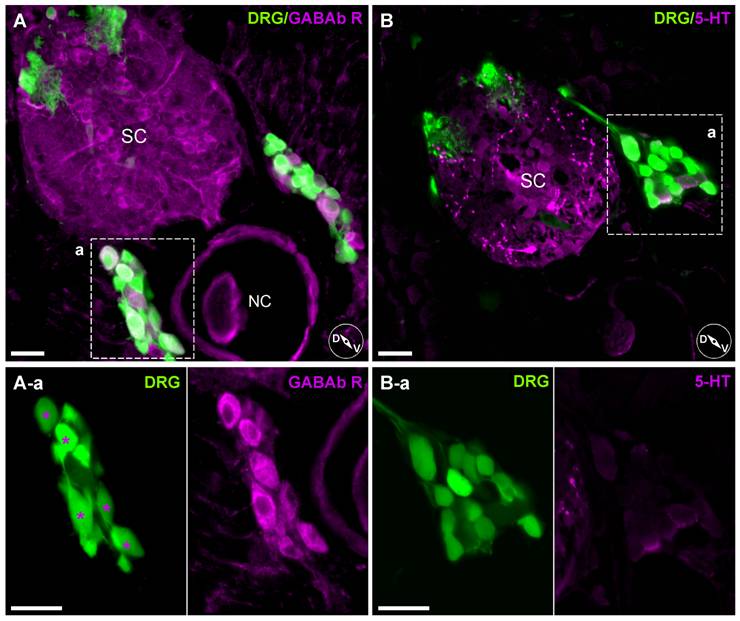
Figure Caption
Fig. 11 Heterogeneous expression pattern of anti-GABAb receptor antibody staining on DRG neurons.
A, Labeling with an anti-GABAb R1 antibody. Enlarged images (A-a, left and right) show only a specific subset of DRG neurons were labeled (asterisks). B, Labeling with an antibody against 5-HT. 5-HT antibody binding was mostly in spinal cord tracts rather than DRG neurons somata (enlarged box B-a). Inset cartoons represent orientation of images. D, dorsal; V, ventral; SC, Spinal Cord; NC, Notochord. Scale bars represent 20 μm.
Acknowledgments
This image is the copyrighted work of the attributed author or publisher, and
ZFIN has permission only to display this image to its users.
Additional permissions should be obtained from the applicable author or publisher of the image.
Full text @ PLoS One