
|
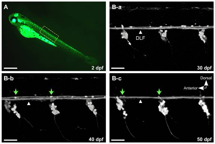
Fig. 1 EGFP expression driven by the Isl2b promoter facilitates observation of dorsal root ganglia development concurrent with degeneration of Rohon-Beard (R-B) neurons.
A, At 2 days post-fertilization (dpf), EGFP was intensely expressed in R-B and DRG neurons. B-a–c, Higher magnification confocal images of the spinal cord in isl2b:EGFP at 30, 40 and 50 dpf. A few EGFP-labeled cells remained over the dorsal longitudinal fasciculus (DFL, white triangles) that progressively decreased until only ~1–3 cells/field remained (B-b and c, green arrow heads). All images are maximum intensity projections of z-stacks acquired in the lateral plane using confocal microscopy. The contrast of the images was adjusted to emphasize EGFP-labeled sensory neurons. Scale bar in A represents 0.5 mm and the other scale bars represent 50 μm.