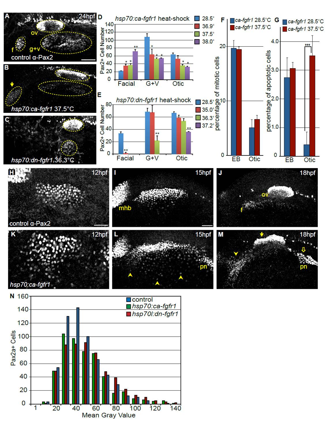
Fig. S7 Modulating Fgf signaling affects size of the otic and EB placodes. (A-C) Representative confocal projections of embryos immunolabeled with anti-Pax2 antibody at 24 hpf including: control (A), constitutively active Fgfr1 Tg(hsp70:ca-fgfr1)pd3 transgene (B) and dominant-negative Fgfr1 Tg(hsp70:dnfgfr1-EGFP)pd1 transgene (C). Heat-shock induction for both constructs was carried out at 10 hpf. Note an increase in the number of Pax2+ cells in the facial placode (arrow in B) and concurrent reduction in the G+V placodes and dysmorphia in the otic vesicle following Fgf upregulation. By contrast, inhibition of Fgf results in loss of the facial placode with a concomitant reduction in the otic vesicle and G+V placodes (C). (D) Quantification of Pax2+ cells in the otic vesicle and EB placodes in control embryos (no heat-shock) and Tg(hsp70:ca-fgfr1)pd3 embryos heat-shocked at various temperatures (36.9, 37.5 and 38°C). Note the 3.4-fold increase in the number of Pax2+ cells in the facial placode following heat-shock at 38°C, with concomitant decrease of the G+V placodes and otic vesicle (Student’s t-test, **P<0.0075). (E) Quantification of Pax2+ cells in the otic vesicle and EB placodes in control embryos (no heat-shock) and Tg(hsp70:dnfgfr1-EGFP)pd1 embryos following heat-shock at various temperatures (35, 36.3 and 37.2°C). Note the <95% reduction in the facial placode following 35°C heat-shock (Student’s t-test, **P<0.002), and complete loss of this placode at higher inductive temperatures. Whereas the G+V placodes are unaffected at lower inductive conditions, there is a 68% reduction (Student’s t-test **P<0.004) and a complete loss of this domain following heat-shock at 36.3°C and 37.2°C, respectively. By contrast, the number of Pax2a+ cells in the otic vesicle is only reduced following heat-shock at the 37.2°C (46% reduction; Student’s t-test **P<0.001). (F) Percentage of Pax2a+ cells undergoing mitosis was measured by immunolabeling with a pH3 antibody at 18 hpf. There is no significant change in proliferation following 10 hpf heat-shock of Tg(hsp70:ca-fgfr1)pd3 embryos, compared with uninduced controls (n=12 embryos per condition). (G) Percentage of Pax2a+ cells undergoing apoptosis as measured by immunolabeling with Caspase-3 antibody at 18 hpf. There is a significant increase of cell death in the otic placode following 10 hpf heat-shock of Tg(hsp70:ca-fgfr1)pd3 embryos (n=12 embryos per condition). (H-M) Pax2a expression at 12, 15 and 18 hpf in control embryos (H-J) or Tg(hsp70:ca-fgfr1)pd3 transgenic embryos following heat-shock induction at 10 hpf (K-M). Note additional Pax2a+ cells lateral to the PPA at 15 hpf (L, arrowheads); and expansion and fusion of Pax2a+ cells in the pronephros (pn) with the PPA. By 18 hpf a striking expansion of the facial placode can be observed (arrowheads), concurrent with a dysmorphic otic vesicle (filled arrow) expansion and mislocalization of the pronephros (M, open arrow). (N) Distribution of Pax2a fluorescence intensity in PPA cells at 12 hpf in control, Tg(hsp70:dnfgfr1-EGFP)pd1 and Tg(hsp70:ca-fgfr1)pd3 following 10 hpf heat- shock. Note that the overall distribution of Pax2a intensity levels is unchanged regardless of Fgf levels (n≥504 cells from eight embryos per condition). Scale bars: 50 μm.
Image
Figure Caption
Acknowledgments
This image is the copyrighted work of the attributed author or publisher, and
ZFIN has permission only to display this image to its users.
Additional permissions should be obtained from the applicable author or publisher of the image.
Full text @ Development

